
Chapter 11: NERVOUS TISSUE
Introduction
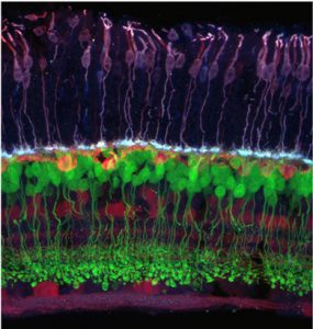
Mouse Retina Cross section of the mouse retina showing the connection between photoreceptor cells (purple) and bipolar neurons (green). [Attribution to R. Wong]
Learning Objectives
Type your learning objectives here.
- Describe the basic structure of a neuron
- Identify the different types of neurons
- Compare the glial cells of the CNS and PNS and describe their function
Nervous tissue is composed of two types of cells, neurons and glial cells. Neurons are responsible for the computation and communication that the nervous system provides. They are electrically active and release chemical signals to target cells. Glial cells, or glia, are known to play a supporting role for nervous tissue.
General Features of Neurons
Neurons are the cells considered to be the basis of nervous tissue. They are responsible for the electrical signals that communicate information about sensations, and that produce movements in response to those stimuli, along with inducing thought processes within the brain. An important part of the function of neurons is in their structure, or shape. The three-dimensional shape of these cells makes the immense numbers of connections within the nervous system possible.
Parts of a Neuron
The main part of a neuron is the cell body, which is also known as the soma (soma = “body”). The cell body contains the nucleus and most of the major organelles. But what makes neurons special is that they have many extensions of their cell membranes, which are generally referred to as processes. Neurons are usually described as having one, and only one, axon—a fibre that emerges from the cell body and projects to target cells. That single axon can branch repeatedly to communicate with many target cells. It is the axon that propagates the nerve impulse, which is communicated to one or more cells. The other processes of the neuron are dendrites, which receive information from other neurons at specialised areas of contact called synapses. The dendrites are usually highly branched processes, providing locations for other neurons to communicate with the cell body. Information flows through a neuron from the dendrites, across the cell body, and down the axon. This gives the neuron a polarity—meaning that information flows in this one direction. Figure 12.8 shows the relationship of these parts to one another.
Where the axon emerges from the cell body, there is a special region referred to as the axon hillock. This is a tapering of the cell body toward the axon fibre.
Many axons are wrapped by an insulating substance called myelin, which is actually made from glial cells. Myelin acts as insulation much like the plastic or rubber that is used to insulate electrical wires. A key difference between myelin and the insulation on a wire is that there are gaps in the myelin covering of an axon. Each gap is called a node of Ranvier and is important to the way that electrical signals travel down the axon. The length of the axon between each gap, which is wrapped in myelin, is referred to as an axon segment. At the end of the axon is the axon terminal, where there are usually several branches extending toward the target cell, each of which ends in an enlargement called a synaptic end bulb. These bulbs are what make the connection with the target cell at the synapse.
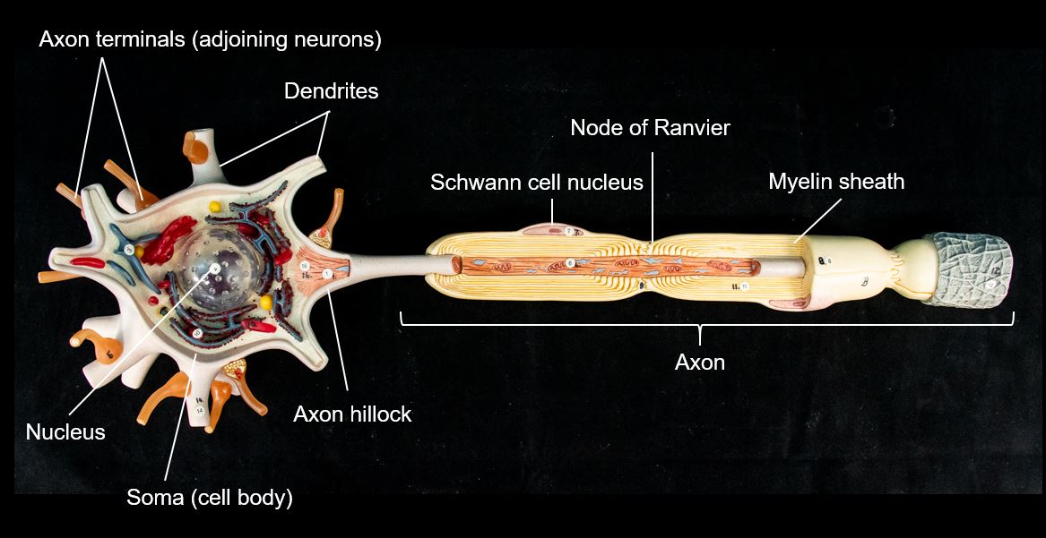
Figure 12.8 Parts of a Neuron The major parts of the neuron are labeled on a multipolar neuron from the PNS.
Types of Neurons
There are many neurons in the nervous system—a number in the trillions. And there are many different types of neurons. They can be classified by many different criteria. The first way to classify them is by the number of processes attached to the cell body. Using the standard model of neurons, one of these processes is the axon, and the rest are dendrites. Because information flows through the neuron from dendrites or cell bodies toward the axon, these names are based on the neuron’s polarity (Figure 12.9)
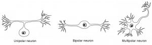
Figure 12.9 Neuron Classification by Shape Unipolar cells have one process that includes both the axon and dendrite. Bipolar cells have two processes, the axon and a dendrite. Multipolar cells have more than two processes, the axon and two or more dendrites.
Neurons can also be classified on the basis of where they are found, who found them, what they do, or even what chemicals they use to communicate with each other. Some neurons referred to in this section on the nervous system are named on the basis of those sorts of classifications (Figure12.10).
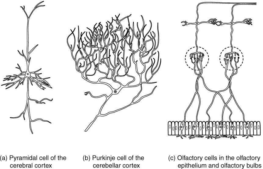
Figure 12.10 Other Neuron Classifications Three examples of neurons that are classified on the basis of other criteria. (a) The pyramidal cell is a multipolar cell with a cell body that is shaped something like a pyramid. (b) The Purkinje cell in the cerebellum was named after the scientist who originally described it. (c) Olfactory neurons are named for the functional group with which they belong.
Glial Cells and Myelination
Glial cells, or neuroglia, are the other type of cell found in nervous tissue. They are considered to be supporting cells, and many functions are directed at helping neurons complete their function for communication. The name glia comes from the Greek word that means “glue”. There are several types of glial cells. Some are found in the central nervous system (brain and spinal cord) and others in the peripheral nervous system (peripheral nerves).
One cell providing support to neurons of the CNS is the astrocyte, so named because it appears to be star-shaped under the microscope (astro- = “star”). Astrocytes have many processes extending from their main cell body (not axons or dendrites like neurons, just cell extensions). Those processes extend to interact with neurons, blood vessels, or the connective tissue covering the CNS that is called the pia mater. Generally, they are supporting cells for the neurons in the central nervous system. Some ways in which they support neurons in the central nervous system are by maintaining the concentration of chemicals in the extracellular space, removing excess signaling molecules, reacting to tissue damage, and contributing to the blood-brain barrier (BBB). The blood-brain barrier is a physiological barrier that keeps many substances that circulate in the rest of the body from getting into the central nervous system, restricting what can cross from circulating blood into the CNS. Nutrient molecules, such as glucose or amino acids, can pass through the BBB, but other molecules cannot.
INTERACTIVE ACTIVITY
Myelin
The insulation for axons in the nervous system is provided by glial cells, oligodendrocytes in the CNS, and Schwann cells in the PNS. Myelin is a lipid-rich sheath that surrounds the axon and by doing so creates a myelin sheath that facilitates the transmission of electrical signals along the axon, ensuring the message is transmitted rapidly along each axon. Myelin is formed by the membrane of the glial cell.
INTERACTIVE ACTIVITY
The glial cell is wrapped around the axon several times with little to no cytoplasm between the glial cell layers. For oligodendrocytes, the rest of the cell is separate from the myelin sheath as a cell process extends back toward the cell body. A few other processes provide the same insulation for other axon segments in the area. For Schwann cells, the outermost layer of the cell membrane contains cytoplasm and the nucleus of the cell as a bulge on one side of the myelin sheath. The inner edge wraps around the axon, creating several layers, and the other edge closes around the outside so that the axon is completely enclosed (Figure12.13a).
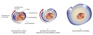
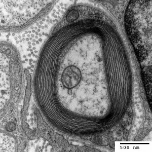
Figure 12.13 The Process of Myelination Myelinating glia wrap several layers of cell membrane around the cell membrane of an axon segment. A single Schwann cell insulates a segment of a peripheral nerve, whereas in the CNS, an oligodendrocyte may provide insulation for a few separate axon segments.
The Synapse
A synapse is a specialised junction mediating the transfer of electrical or chemical information between a neuron and its target effector cell. The synapse is comprised of the axon terminal membrane of the presynaptic neuron (the neuron transmitting the original message), the synaptic cleft (the gap between the two cells), and the cell membrane of the dendrite or cell body of the postsynaptic neuron or the target effector cell (the cell receiving the signal). Chemicals called neurotransmitters are released by the presynaptic neuron and diffuse across the synaptic cleft to bind to receptors on the postsynaptic cell membrane, causing a response in the target effector cell. For example transmission of a message by a second neuron, contraction of a muscle cell, or release of hormones by endocrine cells.
Key Terms
axon hillock tapering of the neuron cell body that gives rise to the axon
axon segment single stretch of the axon insulated by myelin and bounded by nodes of Ranvier at either end (except for the first, which is after the initial segment, and the last, which is followed by the axon terminal)
axon terminal end of the axon, where there are usually several branches extending toward the target cell
bipolar shape of a neuron with two processes extending from the neuron cell body—the axon and one dendrite
central nervous system (CNS) anatomical division of the nervous system located within the cranial and vertebral cavities, namely the brain and spinal cord
dendrite one of many branchlike processes that extends from the neuron cell body and functions as a contact for incoming signals (synapses) from other neurons or sensory cells
glial cell one of the various types of neural tissue cells responsible for maintenance of the tissue, and largely responsible for supporting neurons
multipolar shape of a neuron that has multiple processes—the axon and two or more dendrites
myelin lipid-rich insulating substance surrounding the axons of many neurons, allowing for faster transmission of electrical signals
myelin sheath lipid-rich layer of insulation that surrounds an axon, formed by oligodendrocytes in the CNS and Schwann cells in the PNS; facilitates the transmission of electrical signals
nerve cord-like bundle of axons located in the peripheral nervous system that transmits sensory input and response output to and from the central nervous system
neuron neural tissue cell that is primarily responsible for generating and propagating electrical signals into, within, and out of the nervous system
neurotransmitter chemical signal that is released from the synaptic end bulb of a neuron to cause a change in the target cell
node of Ranvier gap between two myelinated regions of an axon, allowing for strengthening of the electrical signal as it propagates down the axon
nucleus in the nervous system, a localized collection of neuron cell bodies that are functionally related; a “center” of neural function
oligodendrocyte glial cell type in the CNS that provides the myelin insulation for axons in tracts
peripheral nervous system (PNS) anatomical division of the nervous system that is largely outside the cranial and vertebral cavities, namely all parts except the brain and spinal cord
process in cells, an extension of a cell body; in the case of neurons, this includes the axon and dendrites
Schwann cell glial cell type in the PNS that provides the myelin insulation for axons in nerves
soma in neurons, that portion of the cell that contains the nucleus; the cell body, as opposed to the cell processes (axons and dendrites)
synapse narrow junction across which a chemical signal passes from neuron to the next, initiating a new electrical signal in the target cell
synaptic cleft small gap between cells in a chemical synapse where neurotransmitter diffuses from the presynaptic element to the postsynaptic element
synaptic end bulb swelling at the end of an axon where neurotransmitter molecules are released onto a target cell across a synapse
unipolar shape of a neuron which has only one process that includes both the axon and dendrite
