
Chapter 12: NERVOUS SYSTEM
Introduction
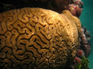
Brain Coral This brain coral appears remarkably similar to its namesake, the brain. Note the extensive ridges (gyri) and valleys (sulci) that increase the surface area of the brain to maximise space for neurons and their connections. [Jaro Nemčok, CC BY-SA 3.0, via Wikimedia Commons]
Chapter Objectives
After studying this chapter, you will be able to:
- Name the major divisions of the nervous system
- Relate the parts of the nervous system to the structure of neurons
- Examine the supporting structures of the central nervous system including the meninges and cerebrospinal fluid
- Explore the major regions of the adult nervous system
- Describe the regions of the spinal cord macroscopically and in cross-section
- Examine the major cranial and spinal nerves
The formation of the nervous system is a miraculous event: Billions of neurons are born and navigate through the developing tissue in the embryo to form meaningful connections with each other and target organs such as muscles. These neurons function in complex ways to allow the body to sense its environment, react to its environment, and integrate information in meaningful ways. The nervous system consists of all nervous tissue components and can be divided into both structural divisions based on the location of this tissue (central or peripheral) and functional divisions based on the type of information being transported (sensory, motor, and autonomic).
Basic Structure and Function of the Nervous System
Learning Objectives
After studying this section, you will be able to:
- Name the major divisions of the nervous system, both anatomical and functional
- Relate the functional and structural differences between gray matter and white matter structures of the nervous system to the structure of neurons
Structural Divisions of the Nervous System
The nervous system can be divided into two major regions based on the location of nervous tissue: the central and peripheral nervous systems. The central nervous system (CNS) is the brain and spinal cord, and the peripheral nervous system (PNS) is everything else (Figure 12.2). The brain is contained within the cranial cavity of the skull, and the spinal cord is contained within the vertebral cavity of the vertebral column.
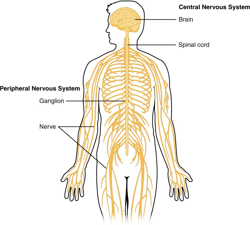
Figure 12.2 Central and Peripheral Nervous System The structures of the PNS are referred to as ganglia and nerves, which can be seen as distinct structures. The main organs of the CNS are the brain and spinal cord.
Functional Divisions of the Nervous System
The nervous system is involved in receiving information about the environment around us (sensation) and generating responses to that information (motor responses). The nervous system can be divided into regions that are responsible for sensation (sensory functions) and for the response (motor functions). But there is a third function that needs to be included. Sensory input needs to be integrated with other sensations, as well as with memories, emotional state, or learning (cognition). Some regions of the nervous system are termed integration or association areas. The process of integration combines sensory perceptions and higher cognitive functions such as memories, learning, and emotion to produce a response.
Controlling the Body
The nervous system can be divided into two parts mostly on the basis of a functional difference in responses. The somatic nervous system (SNS) is responsible for conscious perception and voluntary motor responses. Voluntary motor response means the contraction of skeletal muscle, but those contractions are not always voluntary in the sense that you have to want to perform them. Some somatic motor responses are reflexes, and often happen without a conscious decision to perform them. Other motor responses become automatic (in other words, unconscious) as a person learns motor skills (referred to as “habit learning” or “procedural memory”).
The autonomic nervous system (ANS) is responsible for involuntary control of the body, usually for the sake of homeostasis (regulation of the internal environment). Sensory input for autonomic functions can be from sensory structures tuned to external or internal environmental stimuli. The motor output extends to smooth and cardiac muscle as well as glandular tissue. The role of the autonomic system is to regulate the organ systems of the body, which usually means to control homeostasis. Sweat glands, for example, are controlled by the autonomic system. When you are hot, sweating helps cool your body down. That is a homeostatic mechanism. But when you are nervous, you might start sweating also. That is not homeostatic, it is the physiological response to an emotional state.
Grey and White Matter
Looking at nervous tissue, there are regions that predominantly contain cell bodies and regions that are largely composed of just axons. These two regions within nervous system structures are often referred to as grey matter (the regions with many cell bodies and dendrites) or white matter (the regions with many axons). Figure12.3 demonstrates the appearance of these regions in the brain and spinal cord. The colours ascribed to these regions are what would be seen in “fresh,” or unstained, nervous tissue. Grey matter is not necessarily grey. It can be pinkish because of blood content, or even slightly tan, depending on how long the tissue has been preserved. But white matter is white because axons are insulated by a lipid-rich substance called myelin. Actually, grey matter may have that color ascribed to it because next to the white matter, it is just darker—hence, grey.
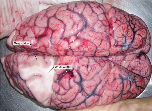
Figure 12.3 Grey Matter and White Matter A brain removed during an autopsy, with a partial section removed, shows white matter surrounded by grey matter. Grey matter makes up the outer cortex of the brain. [modification of work by “Suseno”/Wikimedia Commons]
Regardless of the appearance of stained or unstained tissue, the cell bodies of neurons or axons can be located in discrete anatomical structures that need to be named. Those names are specific to whether the structure is central or peripheral. A localised collection of neuronal cell bodies in the CNS is referred to as a nucleus. In the PNS, a cluster of neuronal cell bodies is referred to as a ganglion. Figure 12.4 indicates how the term nucleus has a few different meanings within anatomy and physiology. It is the centre of an atom, where protons and neutrons are found; it is the centre of a cell, where the DNA is found; and it is a centre of grey matter in the CNS. There is also a potentially confusing use of the word ganglion (plural = ganglia) that has a historical explanation. In the central nervous system, there is a group of nuclei that are connected together and were once called the basal ganglia before “ganglion” became accepted as a description for a peripheral structure. We refer to this group of nuclei as the “basal nuclei” to avoid confusion.
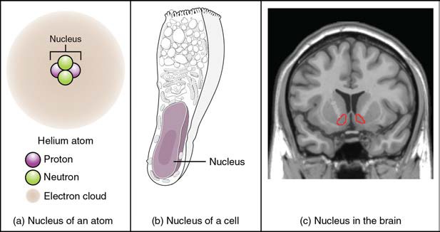
Figure 12.4 What Is a Nucleus? (a) The nucleus of an atom contains its protons and neutrons. (b) The nucleus of a cell is the organelle that contains DNA. (c) A nucleus in the CNS is a localised centre of cell bodies of several neurons, shown here circled in red. [credit c: “Was a bee”/Wikimedia Commons]
Terminology applied to bundles of axons also differs depending on location. A bundle of axons, or fibres, found in the CNS is called a tract whereas the same thing in the PNS would be called a nerve. There is an important point to make about these terms, which is that they can both be used to refer to the same bundle of axons. When those axons are in the PNS, the term is nerve, but if they are CNS, the term is tract. The most obvious example of this is the axons that project from the retina into the brain. Those axons are called the optic nerve as they leave the eye, but when they are inside the cranium, they are referred to as the optic tract. There is a specific place where the name changes, which is the optic chiasm, but they are still the same axons (Figure 12.5). Table 12.1 helps to clarify which of these terms apply to the central or peripheral nervous systems.
Table 12.1 Structures of the CNS and PNS
| Structure | CNS | PNS |
| Group of Neuron Cell Bodies (i.e., gray matter) | Nucleus | Ganglion |
| Bundle of Axons (i.e., white matter) | Tract | Nerve |
Meninges and Flow of Cerebrospinal Fluid
Learning Objectives
After studying this section, you will be able to:
- Describe and identify the meninges and their relationship to the flow of cerebrospinal fluid and blood
- Describe and identify the components of the ventricular system and the regions of the brain in which each is located
- Explain the production of cerebrospinal fluid and its circulation through and around the central nervous system
The CNS is crucial to the operation of the body, and any compromise in the brain and spinal cord can lead to severe difficulties. The CNS has a privileged blood supply, as suggested by the blood-brain barrier. The function of the tissue in the CNS is crucial to the survival of the organism, so the contents of the blood cannot simply pass into the central nervous tissue. To protect this region from the toxins and pathogens that may be travelling through the blood stream, there is strict control over what can move out of the general systems and into the brain and spinal cord. Because of this privilege, the CNS needs specialised structures for the maintenance of circulation. This begins with a unique arrangement of blood vessels carrying fresh blood into the CNS. Beyond the supply of blood, the CNS filters that blood into cerebrospinal fluid (CSF), which is then circulated through the cavities of the brain and spinal cord called ventricles.
Meninges of the Brain and Spinal Cord
The external surface of the CNS is covered by a series of membranes composed of connective tissue called the meninges (pl; meninx, singular), which protect the brain. The meninges are named from external to internal, the dura mater, arachnoid mater and pia mater (Figure 13.17).
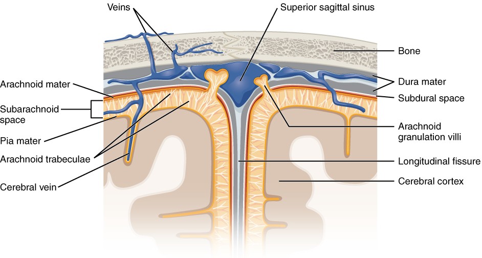
Figure 13.17 Meningeal Layers at the Superior Sagittal Sinus (Coronal View) The layers of the meninges in the longitudinal fissure inferior to the superior sagittal sinus are shown, with the dura mater adjacent to the internal (periosteal) surface of the cranium, the pia mater adjacent to the surface of the brain, and the arachnoid and subarachnoid space between them. An arachnoid villus is shown emerging into the dural sinus to allow CSF to filter back into the blood for drainage.
The same meninges are found supporting the brain and spinal cord. In the spinal cord however the dura mater is not attached to the bony wall of the vertebral canal. Instead there is an adipose tissue filled space called the epidural space between the dura mater and the bone. This is an important location for venous blood flow and is the site for epidural blocks associated with anaesthesia of the body, such as when an expecting mother receives pain relief during childbirth.
The Ventricular System
Cerebrospinal fluid (CSF) circulates throughout and around the CNS. CSF functions to remove metabolic wastes from the interstitial fluids of nervous tissues and return them to the blood. It also acts as a liquid cushion for the brain and spinal cord. The ventricles are the open spaces within the brain where CSF circulates. Cerebrospinal fluid is produced within the ventricles by a type of specialised membrane called a choroid plexus. Ependymal cells (a type of glial cell) surround blood capillaries and filter the blood to make CSF. The fluid is a clear solution with a limited amount of the constituents of blood. It is essentially water, small molecules, and electrolytes. Oxygen and carbon dioxide are dissolved into the CSF, as they are in blood, and can diffuse between the fluid and the nervous tissue. A very small amount of CSF is produced by the choroid plexuses each day, for a total of about 500 milliliters daily, but it is continuously made and pulses through the ventricular system, keeping the fluid moving.
The Ventricles
There are four ventricles within the brain. The first two are named the right and left lateral ventricles and are deep within each cerebral hemisphere. These ventricles are connected to the third ventricle by two openings called the interventricular foramina. The third ventricle is the space between the left and right sides of the diencephalon, which opens into the cerebral aqueduct that passes through the midbrain. The cerebral aqueduct opens into the fourth ventricle, which is the space between the cerebellum, and the pons and superior part of the medulla oblongata (Figure 13.18). The choroid plexuses are found in all four ventricles. The fourth ventricle is continuous with the thin passageway in the spinal cord called the central canal.
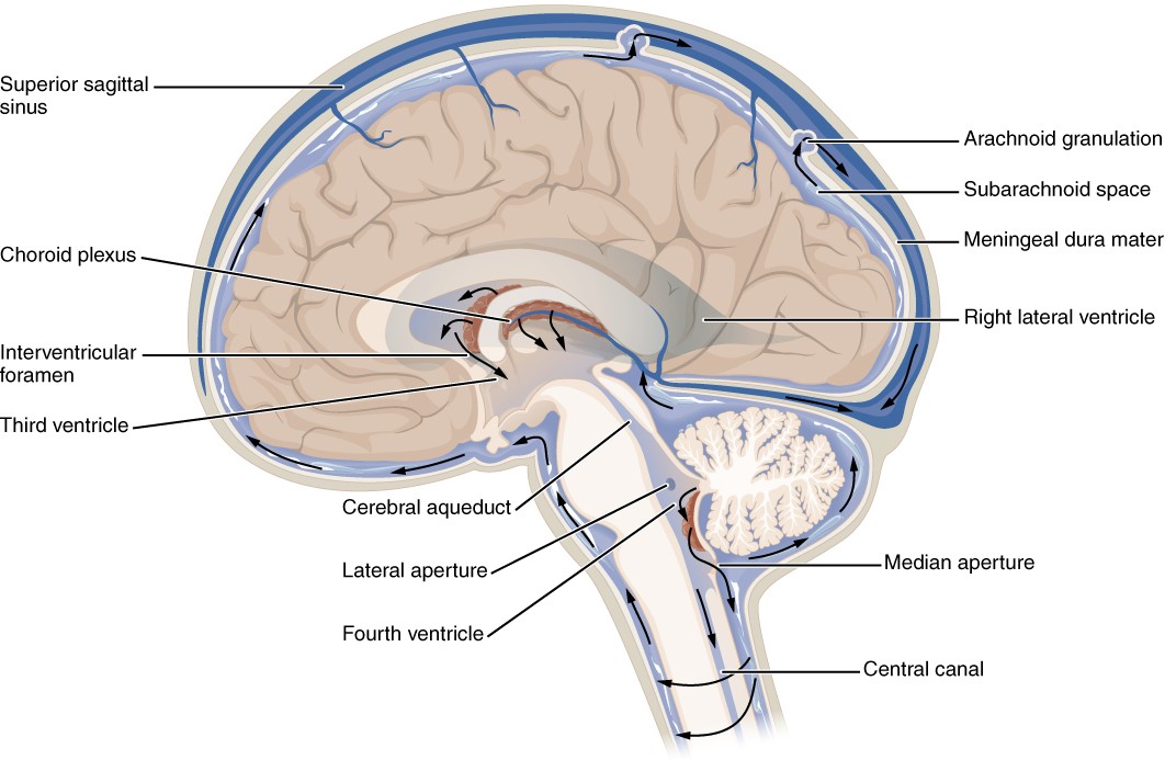
Figure 13.18 Cerebrospinal Fluid Circulation (Midsagittal View). The choroid plexus in the four ventricles produces CSF, which is circulated through the ventricular system and then enters the subarachnoid space through small apertures in the fourth ventricle. The CSF is then reabsorbed into the blood at the arachnoid granulations, where the arachnoid mater protrudes through the dura mater to emerge into the superior sagittal dural sinus.
INTERACTIVE ACTIVITY
Cerebrospinal Fluid Circulation
After flowing through the ventricular system, CSF flows from the fourth ventricle inferiorly into the central canal of the spinal cord as well as posterolaterally through a number of small apertures that open up to the subarachnoid space. The arachnoid villi/granulations are outpocketings of the arachnoid membrane into the dural sinuses so that CSF can be reabsorbed into the blood, along with the metabolic wastes. From the dural sinuses, blood drains out of the head and neck through the internal jugular veins to be reoxygenated by the lungs and wastes to be filtered out by the kidneys.
INTERACTIVE LINK
View this video from Anatomy.TV explaining the flow of cerebrospinal fluid through the ventricles of the brain.
The Central Nervous System: The Brain
Learning Objectives
By the end of this section, you will be able to:
- Describe and identify the major regions and parts of the cerebrum, diencephalon, brainstem and cerebellum
- Describe the connections between the cerebrum and brainstem through the diencephalon
- Explain the arrangement of grey and white matter in the brain
The brain and the spinal cord make up the central nervous system, and they represent the main organs of the nervous system. The spinal cord is a single structure, whereas the adult brain is described in terms of four major regions: the cerebrum, the diencephalon, the brainstem, and the cerebellum.
The Cerebrum
The iconic grey mantle of the human brain, which appears to make up most of the mass of the brain, is the cerebrum (Figure 13.6). The wrinkled external portion composed of grey mattter is the cerebral cortex, and the rest of the structure is deep to that external covering. There is a large separation between the two sides of the cerebrum called the longitudinal fissure. This fissure separates the cerebrum into two distinct halves, a right and left cerebral hemisphere. Deep within the cerebrum, the white matter of the corpus callosum provides the major pathway for axonal communication between the two hemispheres of the cerebral cortex.
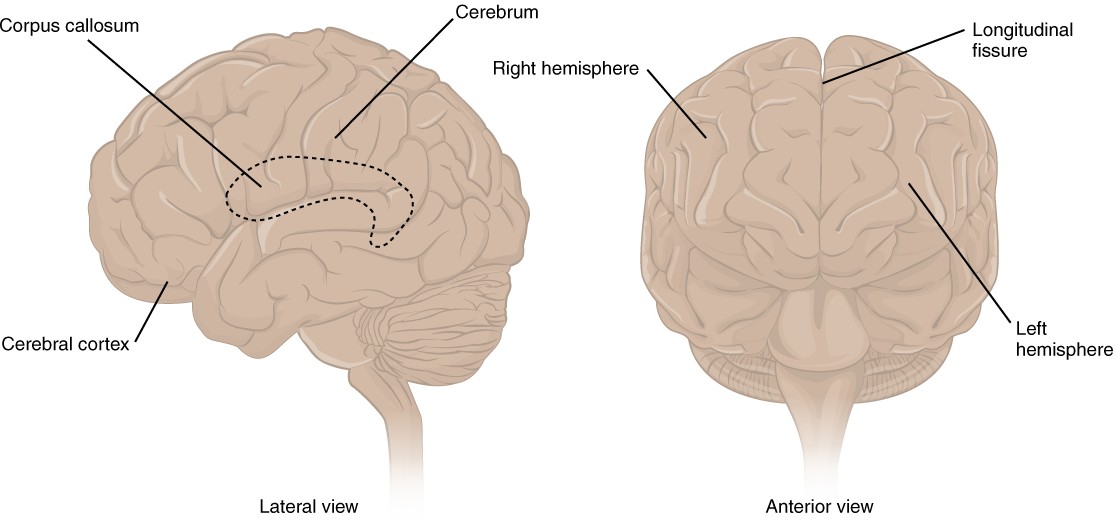
Figure 13.6 The Cerebrum The cerebrum is a large component of the CNS in humans, and the most obvious aspect of it is the folded surface called the cerebral cortex.
Many of the higher neurological functions, such as memory, emotion, and consciousness, are the result of cerebral function. The cerebrum comprises the outer grey matter that is the cortex (from the Latin word meaning “bark of a tree”) and several deep nuclei. The basal nuclei are responsible for cognitive processing, the most important function being that associated with planning movements.
Cerebral Cortex
The cerebrum is covered by a continuous layer of grey matter called the cerebral cortex. This thin, extensive region of wrinkled grey matter is responsible for the higher functions of the nervous system. A gyrus (plural = gyri) is the ridge of one of those wrinkles, and a sulcus (plural = sulci) is the groove between two gyri. The pattern of these folds of tissue indicates specific regions of the cerebral cortex. Extensive folding in the cerebral cortex enables more grey matter to fit into this limited space. If the grey matter of the cortex were peeled off of the cerebrum and laid out flat, its surface area would be roughly equal to one square meter.
The cortex can be separated into four major regions, or lobes (Figure 13.7). The lateral sulcus separates the temporal lobe from the other regions. Superior to the lateral sulcus are the frontal lobe (anterior) and parietal lobe (more posterior), which are separated from each other by the central sulcus. The posterior region of the cortex is the occipital lobe, which has no obvious anatomical border between it and the parietal or temporal lobes on the lateral surface of the brain. From the medial surface, an obvious landmark separates the parietal and occipital lobes and is called the parieto-occipital sulcus.
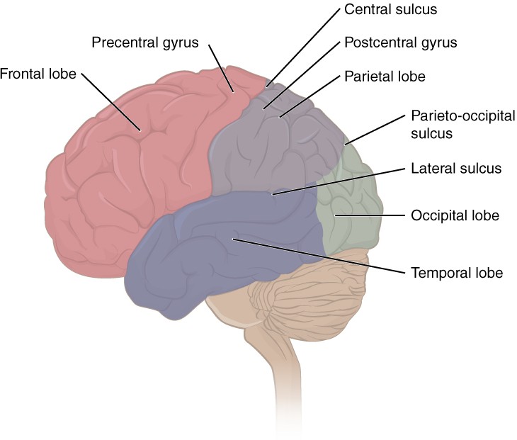
Figure 13.7 Lobes of the Cerebral Cortex The cerebral cortex is divided into four major lobes visible from the external view. Extensive folding increases the surface area available for cerebral functions. Note that the parieto-occipital sulcus is only visible from the medial aspect of the cerebral hemisphere within the longitudinal fissure.
The temporal lobe is associated with primary auditory sensation, memory and smell. The main sensation associated with the parietal lobe is somatosensation, meaning the general sensations associated with the body. Posterior to the central sulcus is the postcentral gyrus, the primary somatosensory cortex. All of the tactile senses are processed in this area, including touch, pressure, tickle, pain, itch and vibration, as well as more general senses of the body such as proprioception and kinesthesia, which are the senses of body position and movement, respectively.
Anterior to the central sulcus is the frontal lobe, which is primarily associated with motor functions. The precentral gyrus is the primary motor cortex. Cells from this region of the cerebral cortex are the upper motor neurons that instruct cells in the spinal cord to move skeletal muscles. Anterior to this region are a few areas that are associated with planned movements. The frontal lobe is also responsible for production of language and cognitive functions that can be the basis of personality, short-term memory and consciousness.
Subcortical structures
The basal nuclei are a set of nuclei (areas of grey matter in the CNS) in the cerebrum (Figure 13.9) responsible for comparing cortical processing with the general state of activity in the nervous system to influence the likelihood of movement taking place. For example, while a student is sitting in a classroom listening to a lecture, the basal nuclei will keep the urge to jump up and scream from actually happening.
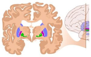
Figure 13.9 Coronal Section of Cerebral Cortex and Basal Nuclei The major components of the basal nuclei, shown in a coronal section of the brain, are coloured blue, purple and green, lying deep in the white matter of the cerebrum. The cerebral cortex can also be seen as the darker brown regions on the most superficial surface of the cerebrum. [By Andrewmeyerson – Own work, CC BY-SA 3.0]
The Diencephalon
The diencephalon is the one region of the adult brain that retains its name from embryologic development. The majority of the brain, the spinal cord, and the PNS all send information to the cerebrum through the diencephalon. Output from the cerebrum passes through the diencephalon. The diencephalon is deep to the cerebrum and constitutes the walls of the third ventricle. The three major regions of the diencephalon are the thalamus centrally, the hypothalamus anteriorly and the epithalamus posteriorly (Figure 13.11). The epithalamus contains the pineal gland, which is an endocrine gland that produces the hormone melatonin important in sleep-wake cycles.
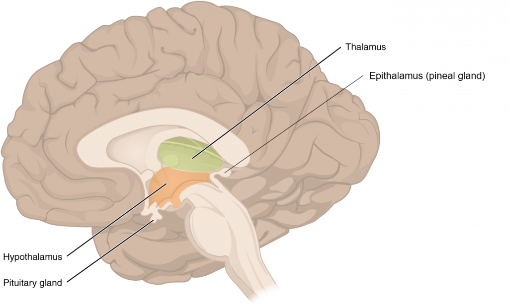
Figure 13.11 The Diencephalon (Midsagittal view of Brain) The diencephalon is composed primarily of the thalamus, hypothalamus and epithalamus, which together define the walls of the third ventricle. The thalami are two elongated, ovoid structures on either side of the midline that make contact in the middle. The hypothalamus is inferior and anterior to the thalamus, culminating in a sharp angle to which the pituitary gland is attached. The epithalamus is the most posterior part of the diencephalon consisting largely of the pineal gland. [Modified from ‘Anatomy and Physiology’, OpenStax]
Brainstem
The midbrain, pons and medulla oblongata are collectively referred to as the brainstem (Figure 13.12). The structure connects the brain to the spinal cord. Posteriorly attached to the brainstem, but considered a separate region of the adult brain, is the cerebellum. The midbrain coordinates sensory representations of the visual, auditory, and somatosensory perceptual spaces. The pons is the main connection with the cerebellum. The pons and the medulla oblongata regulate several crucial functions, including the cardiovascular and respiratory systems and rates.
The cranial nerves connect through the brainstem and provide the brain with the sensory input and motor output associated with the head and neck, including most of the special senses. The major ascending and descending neuronal tracts between the spinal cord and brain, specifically the cerebrum, pass through the brainstem.
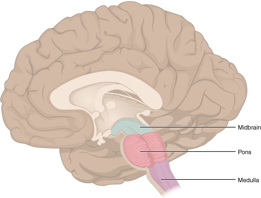
Figure 13.12 The Brainstem (Midsagittal view of Brain) The brainstem comprises three regions: the midbrain (superior), the pons, and the medulla oblongata (inferior).
The Cerebellum
The cerebellum, as the name suggests, is the “little brain” and looks like a miniature version of that part of the brain (Figure 13.13). The cerebellum is largely responsible for comparing information from the cerebrum with sensory feedback from the periphery through the spinal cord. It accounts for approximately 10 percent of the mass of the brain.
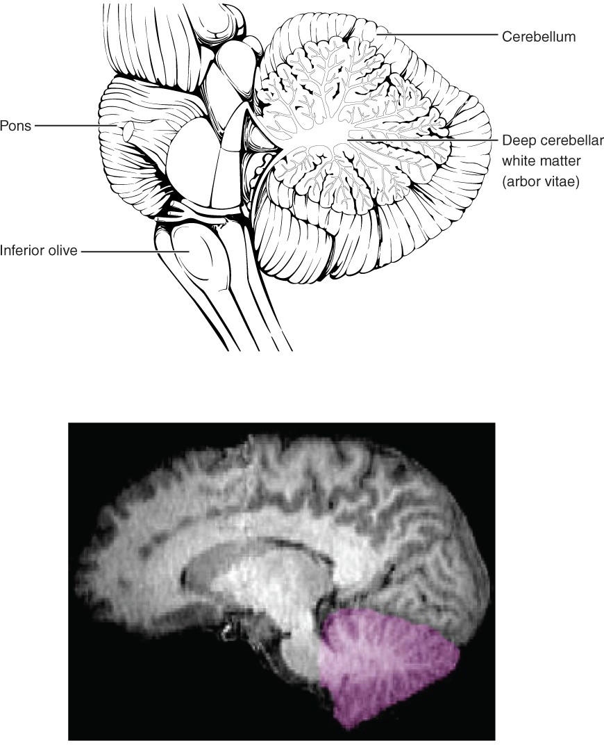
Figure 13.13 The Cerebellum The cerebellum is situated on the posterior surface of the brainstem.
If the primary motor cortex of the frontal lobe sends a command down to the spinal cord to initiate walking, a copy of that instruction is sent to the cerebellum. Sensory feedback from the muscles and joints, proprioceptive information about the movements of walking, and sensations of balance are sent to the cerebellum and the cerebellum compares them. If walking is not coordinated, perhaps because the ground is uneven or a strong wind is blowing, then the cerebellum sends out a corrective command to compensate for the difference between the original cortical command and the sensory feedback. The output of the cerebellum then sends signals into the spinal cord to correct the messages going to skeletal muscles.
INTERACTIVE ACTIVITY
The Central Nervous System: The Spinal Cord
Learning Objectives
By the end of this section, you will be able to:
- Describe and identify the major macroscopic features of the spinal cord
- Describe the connections between the cerebrum and the spinal cord
- Explain the arrangement of grey and white matter in the spinal cord
Macroscopic Anatomy of Spinal Cord
The length of the spinal cord is divided into regions that correspond to the regions of the vertebral column. The name of a spinal cord region corresponds to the same level at which spinal nerves pass through the intervertebral foramina. Immediately adjacent to the brainstem is the cervical region, followed by the thoracic, then the lumbar, and finally the sacral region. The spinal cord is not the full length of the vertebral column because the spinal cord does not grow significantly longer after the first or second year, but the skeleton continues to grow. Despite this difference in length, the nerves that emerge from the spinal cord still pass through the intervertebral foramina at their respective levels. As the vertebral column grows, these nerves grow with it and result in a long bundle of nerves that resembles a horse’s tail and is named the cauda equina. The sacral spinal cord is at the level of the upper lumbar vertebral bones. The spinal nerves extend from their various levels to the proper level of the vertebral column.
Due to the high number of neurons required to innervate the upper and lower limbs, the spinal cord is larger in the cervical and lumbar regions to allow for more room for the cell bodies of the neurons to reside. This results in a noticeable bulging of the spinal cord as the cervical enlargement (associated with the brachial plexus and upper limb innervation) and lumbar enlargement (associated with the lumbosacral plexuses and lower limb innervation). The terminal end of the spinal cord is referred to as the conus medullaris and tapers inferiorly to a point. The conus medullaris is located typically at the vertebral level of L1/L2 in adults. Extending from the conus medullaris is the filum terminale composed of pia mater, which attaches to the coccyx to anchor the spinal cord. The filum terminale can be identified as a white filament in the centre of the nerves that make up the cauda equina. Another structure composed of pia mater is the ligamentum denticulatum that lies lateral to the spinal cord. As its name suggests this structure resembles teeth extending out from the lateral margin of the spinal cord to pierce through the arachnoid mater to attach to the dura mater to support and limit mobility of the spinal cord in the cerebrospinal fluid filled subarachnoid space.
Cross-sectional anatomy of spinal cord
As shown in Figure 13.14, the grey matter of the spinal cord in cross-section is subdivided into regions that are referred to as horns. The posterior (dorsal) horn is responsible for sensory processing. The anterior (ventral) horn sends out motor signals to the skeletal muscles. The dorsal and ventral horns are largest in the cervical and lumbosacral regions of the spinal cord where additional neurons are needed to innervate the upper and lower limbs. The lateral horn, which is only found in the thoracic and upper lumbar regions, is the central component of the sympathetic division of the autonomic nervous system.
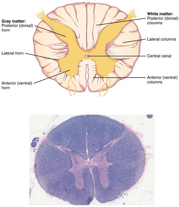
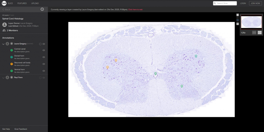
Figure 13.14 Cross-section of Spinal Cord The cross-section of a thoracic spinal cord segment shows the dorsal, ventral and lateral horns of grey matter. LM ×40. (Micrograph provided by the Regents of University of Michigan Medical School © 2012)
Just as the grey matter is separated into horns, the white matter of the spinal cord is separated into columns. Ascending tracts of nervous system fibres in these columns carry sensory information up to the brain, whereas descending tracts carry motor commands from the brain.
An example of a descending tract is the corticospinal tract responsible for the conscious or voluntary movements of skeletal muscles. Any motor command from the primary motor cortex is sent down the axons of motor neurons to reach either the cranial motor nuclei or the ventral horn of the spinal cord. The axons of the corticospinal tract are largely contralateral, meaning that they cross the midline of the brainstem or spinal cord and synapse on the opposite side of the body. Therefore, the right motor cortex of the cerebrum controls muscles on the left side of the body, and vice versa.
An example of an ascending tract is the spinothalamic tract responsible for pain and temperature sensations. The spinothalamic tract begins with neurons in a dorsal root ganglion that have nerve endings in the skin. These neurons extend their axons to the dorsal horn, where they synapse with a second neuron. The name “spinothalamic” comes from this second neuron, which has its cell body in the spinal cord gray matter and connects to the thalamus. Axons from these second neurons then decussate (cross) within the spinal cord and ascend to the brain and enter the contralateral thalamus. The neurons in the thalamus then project their axons to the postcentral gyrus of the cerebral cortex for somatosensory perception.
INTERACTIVE LINK
The Peripheral Nervous System
Learning Objectives
By the end of this section, you will be able to:
- Describe the structures found in the PNS
- Distinguish between somatic and autonomic structures
- Name examples of the cranial nerves and explain the functions associated with each
- Describe the sensory and motor components of spinal nerves and the plexuses that they pass through
Nerves
Bundles of axons in the PNS are referred to as nerves (referred to as tracts in the CNS). Nerves are composed of more than just nervous tissue. They have connective tissues invested in their structure, as well as blood vessels supplying the tissues with nourishment. Nerves are associated with the region of the CNS to which they are connected, either as cranial nerves connected to the brain or spinal nerves connected to the spinal cord.
Cranial Nerves
The nerves attached to the brain are the cranial nerves, which are primarily responsible for the sensory and motor functions of the head and neck. A number of cranial nerves are also responsible for parasympathetic innervation of visceral organs such as the organs of the thoracic and abdominal cavities. There are twelve cranial nerves, which are designated CNI through CNXII for “Cranial Nerve,” using Roman numerals 1 through 12 (Figure 13.23). They can be classified as sensory nerves, motor nerves, or a combination of both (mixed nerves). The names of the cranial nerves are listed in Table 13.3 along with a brief description of their function, their source (sensory ganglion or motor nucleus), and their target (sensory nucleus or skeletal muscle).
The olfactory nerve and optic nerve are responsible for the sense of smell and vision, respectively. The oculomotor nerve is responsible for eye movements by controlling four of the extraocular muscles. It is also responsible for lifting the upper eyelid when the eyes point up, and for pupillary constriction. The trochlear nerve and the abducens nerve are both responsible for eye movement, but do so by controlling different extraocular muscles. The trigeminal nerve is responsible for cutaneous sensations of the face and controlling the muscles of mastication. The facial nerve is responsible for the muscles involved in facial expression, as well as part of the sense of taste and the production of saliva. The vestibulocochlear nerve is responsible for the senses of hearing and balance. The glossopharyngeal nerve is responsible for controlling muscles in the oral cavity and superior pharynx, as well as part of the sense of taste and the production of saliva. The vagus nerve is responsible for contributing to homeostatic control of the organs of the thoracic and superior abdominal cavities. The accessory nerve is responsible for controlling the muscles of the neck, along with cervical spinal nerves. The hypoglossal nerve is responsible for controlling the muscles of the inferior pharynx and tongue.
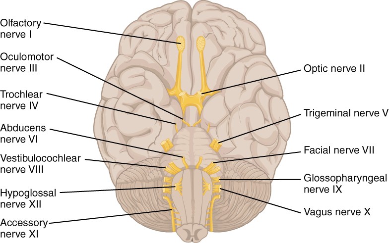
Figure 13.23 The Cranial Nerves The anatomical arrangement of the roots of the cranial nerves observed from an inferior view of the brain.
Three of the cranial nerves also contain autonomic fibres, and a fourth is almost purely a component of the autonomic system. The oculomotor, facial and glossopharyngeal nerves contain fibres that contact autonomic ganglia. The oculomotor fibres initiate pupillary constriction, whereas the facial and glossopharyngeal fibres both initiate salivation. The vagus nerve primarily targets autonomic ganglia in the thoracic and superior abdominal cavities to slow down heart rate and breathing, and activate digestive function.
Another important aspect of the cranial nerves is the functional role each nerve plays. The first, second, and eighth nerves are purely sensory: the olfactory (CNI), optic (CNII), and vestibulocochlear (CNVIII) nerves. The three eye-movement nerves are all motor: the oculomotor (CNIII), trochlear (CNIV), and abducens (CNVI). The spinal accessory (CNXI) and hypoglossal (CNXII) nerves are also strictly motor. The remainder of the nerves contain both sensory and motor fibres. They are the trigeminal (CNV), facial (CNVII), glossopharyngeal (CNIX), and vagus (CNX) nerves.
Table 13.3 Cranial Nerves
|
# |
Name |
Function (S/M/B) |
Parasympathetic function |
|
I |
Olfactory |
Smell (S) |
N/A |
|
II |
Optic |
Vision (S) |
N/A |
|
III |
Oculomotor |
Eye movements (inferior oblique, medial rectus, superior rectus, inferior rectus) (M) |
Pupillary reflex (adjusts size of pupil to change amount of light entering eye) and accommodation reflex (adjusts lens to focus on near/far objects) of eye |
|
IV |
Trochlear |
Eye movements (superior oblique) (M) |
N/A |
|
V |
Trigeminal |
Sensory of face / motor – muscles of mastication (B) |
N/A |
|
VI |
Abducens |
Eye movements (lateral rectus) (M) |
N/A |
|
VII |
Facial |
Motor – muscles of facial expression (B) |
Salivation |
|
VIII |
Auditory (Vestibulocochlear) |
Hearing/ balance (S) |
N/A |
|
IX |
Glossopharyngeal |
Motor – pharynx (B) |
Salivation |
|
X |
Vagus |
Motor (muscles of larynx and neck) / sensory (B) |
Heart rate, breathing, digestion |
|
XI |
Accessory |
Motor – head and neck (M) |
N/A |
|
XII |
Hypoglossal |
Motor – inferior pharynx (M) |
N/A |
Spinal Nerves
The nerves connected to the spinal cord are the spinal nerves. The arrangement of these nerves is much more regular than that of the cranial nerves. All of the spinal nerves are combined sensory and motor axons that separate into two nerve roots. The sensory axons enter the spinal cord as the dorsal nerve root. The motor fibres, both somatic and autonomic, emerge as the ventral nerve root. The dorsal root ganglion for each nerve is an enlargement of the dorsal root of the spinal nerve where the cell bodies of sensory pseudounipolar neurons are housed. Lateral to the dorsal root ganglion the dorsal and ventral nerve roots unite to form the body of the spinal nerve. Soon after this union the spinal nerve then divides into a dorsal ramus (singular; rami = plural) and ventral ramus of the spinal nerve. The spinal nerve body and rami are mixed fibres containing both motor and sensory neurons. The dorsal ramus of the spinal nerve is the smaller of the two rami and innervates largely organs of the back. The ventral ramus of the spinal nerve is the larger branch of the body of the spinal nerve and innervates most regions of the body anterior to the vertebral column.
INTERACTIVE ACTIVITY
Review the parts of a spinal nerve and its relationship to the spinal cord at the cervical vertebra level in this series of photos of a plastic model. This activity requires you to move from one slide to the next. You can do this by clicking either the arrows beside the image, or the buttons below the image. Test your knowledge on the unlabelled photo before revealing the names of the main parts in the labelled photo.
Questions:
(a) Name the space that the body of the spinal nerve passes through between adjacent vertebrae
(b) Why is the ventral ramus so much bigger than the dorsal ramus?
There are 31 spinal nerves, named for the level of the spinal cord at which each one emerges. There are eight pairs of cervical nerves designated C1 to C8, twelve thoracic nerves designated T1 to T12, five pairs of lumbar nerves designated L1 to L5, five pairs of sacral nerves designated S1 to S5, and one pair of coccygeal nerves. The nerves are numbered from the superior to inferior positions, and each emerges from the vertebral column through the intervertebral foramen at its level. The first nerve, C1, emerges between the first cervical vertebra and the occipital bone. The second nerve, C2, emerges between the first and second cervical vertebrae. Spinal nerves C2 to C7 emerge superior to the vertebra of the same name, but C8 emerges between the seventh cervical vertebra and the first thoracic vertebra. For the thoracic and lumbar nerves, each one emerges inferior to the vertebra that has the same designation. The sacral nerves emerge through the sacral foramina along the length of the sacrum.
Spinal nerves extend laterally away from the vertebral column to innervate the periphery. Axons from different spinal nerves will come together into a peripheral nerve. Most nerves in the periphery are not straight continuations of the spinal nerves, but rather the reorganisation of the axons in the ventral spinal rami to follow different courses. This occurs at four places along the length of the vertebral column, each identified as a network of nerve fibres called a nerve plexus. Of the four nerve plexuses, two are found at the cervical level, one at the lumbar level, and one at the sacral level (Figure 13.24). Spinal nerves of the thoracic region, T2 through T11, are not part of the plexuses but rather emerge and give rise to the intercostal nerves found between the ribs.
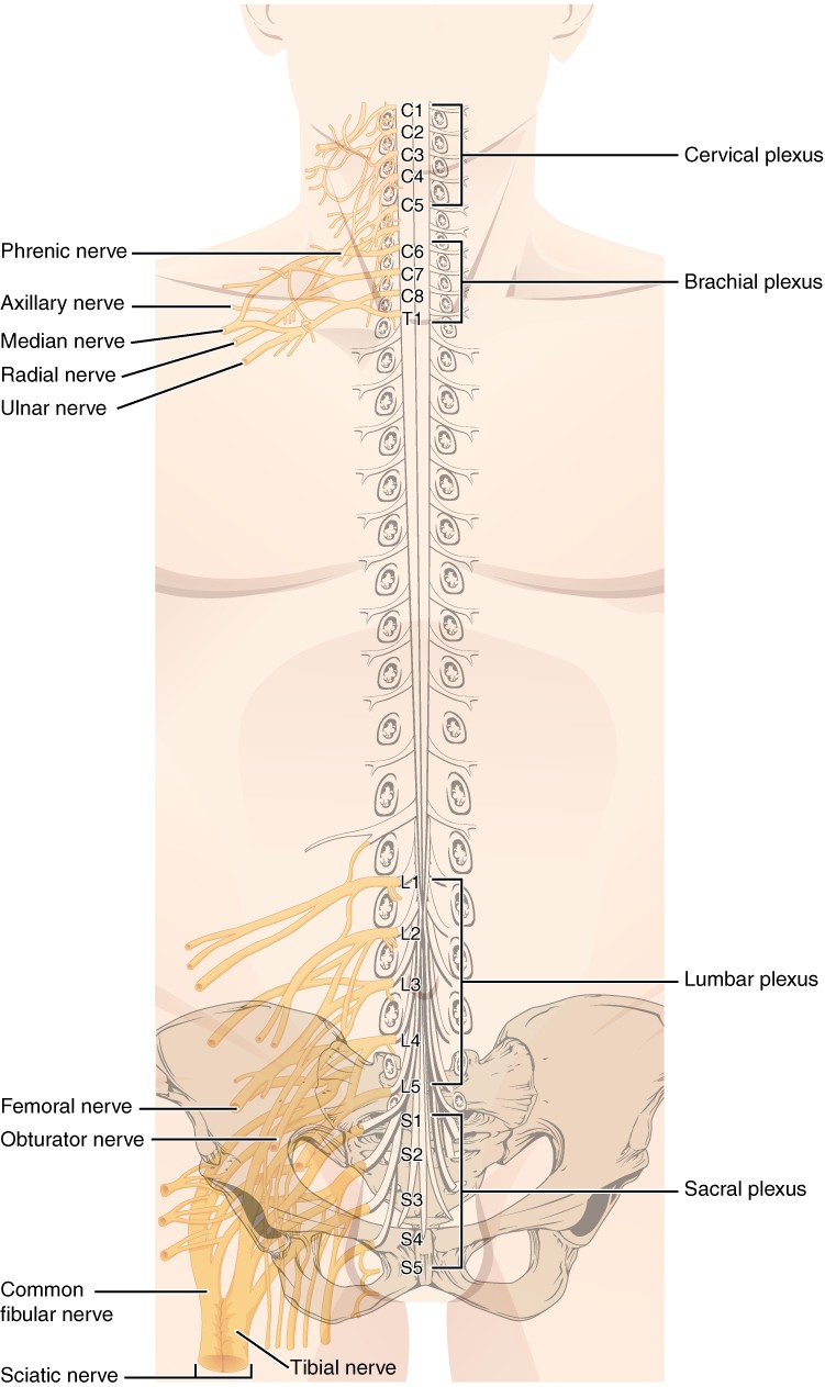
Figure 13.24 Nerve Plexuses of the Body There are four main nerve plexuses in the human body. The cervical plexus supplies nerves to the posterior head and neck, as well as to the thoracic diaphragm. The brachial plexus supplies nerves to the upper limb. The lumbar plexus supplies nerves to the anterior parts of the lower limb. The sacral plexus supplies nerves to the posterior parts of the lower limb.
Autonomic Nervous System
Learning Objectives
By the end of this section, you will be able to:
- Describe the major function of the autonomic nervous system
- Describe and identify the major parts of the autonomic nervous system in the PNS
- Describe the connection between autonomic structures in the CNS and PNS
The structures and function of the autonomic nervous system have been referred to throughout earlier sections of this chapter. To recap, the autonomic nervous system (ANS) is responsible for involuntary control of the body, usually for the sake of homeostasis (regulation of the internal environment). Sensory input for autonomic functions can be from sensory structures tuned to external or internal environmental stimuli. The motor output extends to smooth and cardiac muscle as well as glandular tissue. The role of the autonomic system is to regulate the organ systems of the body, which usually means to control homeostasis.
Autonomic nerve fibres travel through both the central and peripheral nervous systems. Its basic unit consists of a series of two motor neurons which synapse in a ganglion (collection of neuronal cell bodies in the PNS); these neurons are referred to as the preganglionic and postganglionic neurons.
There are two subdivisions of the autonomic nervous system, namely the sympathetic nervous system and the parasympathetic nervous system. Cardiac muscle, smooth muscle of tubular organs and most glands are innervated by both subdivisions. For each tissue or gland one subdivision of the ANS increases its activity (excitatory function) whereas the other subdivbision of the ANS decreases its activity (inhibitory function). The activity of cardiac muscle is increased by sympathetic innervation and decreased by parasympathetic innervation. The parasympathetic system increases the activity of smooth muscle of the intestinal wall whereas the sympathetic system decreases its activity to redirect blood flow towards the skeletal muscles. Sweat glands, vessels and suprarenal/adrenal glands are innervated solely by the sympathetic nervous system. The combined functions of each subdivision of the ANS regulate the body in response to contrasting stimuli. The parasympathetic nervous system function is referred to as rest and digest, in contrast to the sympathetic nervous system function being referred to as fight or flight.
Key Terms
abducens nerve sixth cranial nerve; responsible for contraction of one of the extraocular muscles
accessory nerve eleventh cranial nerve; responsible for contraction of neck muscles
arachnoid granulation outpocket of the arachnoid membrane into the dural sinuses that allows for reabsorption of CSF into the blood; granulation refers to the hardened adult structure compared to arachnoid villi which refer to the soft structure found in children
arachnoid mater middle layer of the meninges named for the spider-web–like trabeculae that extend between it and the pia mater
ascending spinal tract central nervous system fibres carrying sensory information from the spinal cord or periphery to the brain
basal nuclei nuclei of the cerebrum (with a few components in the superior brainstem and diencephalon) that are responsible for assessing cortical movement commands and comparing them with the general state of the individual; largely related to motor functions, as evidenced through the symptoms of Parkinson’s and Huntington’s diseases
brachial plexus nerve plexus associated with the inferior cervical spinal nerves and first thoracic spinal nerve
brainstem region of the adult brain that includes the midbrain, pons, and medulla oblongata
cauda equina bundle of spinal nerve roots that descend from the inferior spinal cord inferior to the first lumbar vertebra and lie within the vertebral cavity; has the appearance of a horse’s tail
central canal hollow space within the spinal cord that contains cerebrospinal fluid
central sulcus surface landmark of the cerebral cortex that marks the boundary between the frontal and parietal lobes
cerebellum region of the adult brain connected primarily to the pons and is largely responsible for comparing information from the cerebrum with sensory feedback from the periphery through the spinal cord
cerebral aqueduct connection of the ventricular system between the third and fourth ventricles located in the midbrain
cerebral cortex external grey matter covering the cerebrum, marked by ridges and valleys known as gyri and sulci, respectively
cerebral hemisphere one half of the bilaterally symmetrical cerebrum
cerebrum region of the adult brain responsible for higher neurological functions such as memory, emotion, and consciousness
cervical plexus nerve plexus associated with the superior cervical spinal nerves
choroid plexus specialised structures containing ependymal cells lining blood capillaries that filter blood to produce CSF in the four ventricles of the brain
corpus callosum large white matter structure that allows axons of neurons to cross between the right and left cerebral hemispheres
cranial nerve one of twelve nerves connected to the brain that are responsible for sensory or motor functions of the head and neck
descending tract central nervous system fibres carrying motor commands from the brain to the spinal cord or periphery
diencephalon region of the adult brain that retains its name from embryonic development and includes the thalamus, hypothalamus and epithalamus
dorsal (posterior) horn gray matter region of the spinal cord in which sensory input arrives, sometimes referred to as the dorsal horn
dorsal (posterior) nerve root axons entering the posterior horn of the spinal cord
dorsal (posterior) root ganglion sensory ganglion attached to the posterior nerve root of a spinal nerve; contains the cell bodies of sensory neurons
dura mater tough, fibrous, outer layer of the meninges that is attached to the inner surface of the cranium and vertebral column and surrounds the entire CNS
dural sinus any of the venous structures surrounding the brain, enclosed within the dura mater, which drain blood from the CNS to the common venous return of the jugular veins
epithalamus region of the diencephalon containing the pineal gland
extraocular muscles six skeletal muscles that control eye movement within the orbit
facial nerve seventh cranial nerve; responsible for contraction of the muscles of facial expression and for part of the sense of taste, as well as causing saliva production
femoral nerve systemic nerve of the anterior leg that arises from the lumbar plexus
foramen magnum large opening in the occipital bone of the skull through which the spinal cord emerges
fourth ventricle the portion of the ventricular system that is in the region of the brainstem and opens into the subarachnoid space through apertures
frontal lobe most anterior region of the cerebral cortex responsible for voluntary motor movement, personality, planning and intelligence
glossopharyngeal nerve ninth cranial nerve; responsible for contraction of muscles in the tongue and pharynx and for part of the sense of taste, as well as causing saliva production
gyrus ridge formed by convolutions on the surface of the cerebrum
hypoglossal nerve twelfth cranial nerve; responsible for contraction of muscles of the tongue
hypothalamus major region of the diencephalon that is responsible for coordinating autonomic and endocrine control of homeostasis
intercostal nerve systemic nerve in the thoracic cavity that is found between two ribs
interventricular foramina openings between the lateral ventricles and third ventricle allowing for the passage of CSF
kinesthesia general sensory perception of movement of the body
lateral horn region of the spinal cord grey matter in the thoracic and upper lumbar regions that is the central component of the sympathetic division of the autonomic nervous system
lateral sulcus surface landmark of the cerebral cortex that marks the boundary between the temporal lobe and the frontal and parietal lobes
lateral ventricles bilateral portions of the ventricular system that are in the region of the cerebral hemispheres
longitudinal fissure large separation along the midline between the two cerebral hemispheres
lumbar plexus nerve plexus associated with the lumbar spinal nerves
meninges protective outer coverings of the CNS composed of connective tissue
midbrain middle region of the adult brainstem
nerve plexus network of nerves in the PNS
occipital lobe region of the cerebral cortex responsible for vision perception
oculomotor nerve third cranial nerve; responsible for contraction of four of the extraocular muscles, the muscle in the upper eyelid, and pupillary constriction and the accommodation reflex
olfaction special sense responsible for smell, which has a unique, direct connection to the cerebrum
olfactory nerve first cranial nerve; responsible for the sense of smell
optic nerve second cranial nerve; responsible for visual sensation
parietal lobe region of the cerebral cortex responsible for somatosensation
parieto-occipital sulcus groove in the medial surface of the cerebral cortex representing the border between the parietal and occipital cortices
peripheral nerve nerve in the periphery distal to a nerve plexus or spinal nerve
phrenic nerve systemic nerve from the cervical plexus that innervates the thoracic diaphragm
pia mater thin, innermost membrane of the meninges that directly covers the surface of the CNS
postcentral gyrus ridge just posterior to the central sulcus, in the parietal lobe, where somatosensory processing initially takes place in the cerebrum
precentral gyrus primary motor cortex located in the frontal lobe of the cerebral cortex
proprioception general sensory perceptions providing information about location and movement of body parts; the “sense of the self”
sacral plexus nerve plexus associated with the inferior lumbar and sacral spinal nerves
sciatic nerve systemic nerve from the sacral plexus and extends across the hip joint and gluteal region into the posterior femoral region
somatosensation general senses related to the body, usually thought of as the senses of touch, which would include pain, temperature, and proprioception
spinal nerve one of 31 nerves connected to the spinal cord
subarachnoid space space between the arachnoid mater and pia mater that contains CSF and the fibrous connections of the arachnoid trabeculae
sulcus groove formed by convolutions in the surface of the cerebral cortex
superior sagittal sinus dural sinus that runs along the superior border of the longitudinal fissure and drains blood from the majority of the external cerebrum
sympathetic ganglia autonomic ganglia in a chain along the anterolateral aspect of the vertebral column that are responsible for contributing to homeostatic mechanisms of the autonomic nervous system
temporal lobe region of the cerebral cortex responsible for processing of sound, memory, smell and emotion
thalamus major region of the diencephalon that is responsible for relaying information between the cerebrum and the brainstem, spinal cord, and periphery
third ventricle portion of the ventricular system that is in the region of the diencephalon
trigeminal nerve fifth cranial nerve; responsible for cutaneous sensation of the face and contraction of the muscles of mastication
trochlear nerve fourth cranial nerve; responsible for contraction of one of the extraocular muscles
vagus nerve tenth cranial nerve; responsible for the autonomic control of organs in the thoracic and upper abdominal cavities
ventral horn gray matter of the spinal cord containing multipolar motor neurons, sometimes referred to as the anterior horn
ventricles spaces for cerebrospinal fluid to circulate through the brain
vestibulocochlear nerve eighth cranial nerve; responsible for the sensations of hearing and balance
