
Chapter 4: EPITHELIAL TISSUE
Introduction
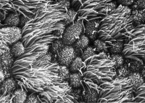
Uterine tube epithelium A scanning electron micrograph showing the ciliated and non-ciliated cells comprising the simple columnar epithelium of the uterine (Fallopian) tube. [https://embryology.med.unsw.edu.au/embryology/index.php?curid=6801]
Learning Objectives
By the end of this section, you will be able to:
- Describe the structural features, location and major functions of surface epithelial tissue
- Identify in photomicrographs simple epithelia and stratified epithelia, including squamous, cuboidal, and columnar epithelia subtypes
- Identify examples and describe the structure and secretions of endocrine and exocrine glands
Most epithelial tissues are essentially large sheets of cells covering all the surfaces of the body exposed to the outside world and lining the outside of organs. Epithelium also forms much of the glandular tissue of the body. Skin is not the only area of the body exposed to the external environment. Other areas include the airways, the digestive tract, as well as the urinary and reproductive systems, all of which are lined by an epithelium. Hollow organs and body cavities that do not connect to the exterior of the body, which includes, blood vessels and serous membranes, are lined by endothelium (plural = endothelia), which is a type of epithelium.
All epithelial tissue is highly cellular, with little or no extracellular material present between cells. Adjoining cells form a specialised intercellular connection between their cell membranes called a cell junction. The epithelial cells exhibit differences in structure and function between the exposed or apical facing surface of the cell and the basal surface close to the underlying body structures. The basal region of each epithelium attaches to a non-cellular layer called the basement membrane, which separates the epithelium form the underlying connective tissue. Epithelial tissues are nearly completely avascular.
The Epithelial Cell
Epithelial cells are typically characterised by the polarised distribution of organelles and membrane-bound proteins between their basal and apical surfaces. Particular structures found in some epithelial cells are an adaptation to specific functions. Certain organelles are segregated to the basal sides, whereas other organelles and extensions, such as cilia, when present, are on the apical surface.
Classification of Surface Epithelial Tissues
Epithelial tissues can be grouped into two major types: surface epithelia that line internal surfaces and cover external surfaces of the body; and glandular epithelia that form all glands in the body.
Surface epithelial tissues are classified according to the shape of the cells and number of the cell layers formed (Figure 4.5). Cell shapes can be squamous (flattened and thin), cuboidal (boxy, as wide as it is tall), or columnar (rectangular, taller than it is wide). Similarly, the number of cell layers in the tissue can be one—where every cell rests on the basal lamina—which is a simple epithelium, or more than one, which is a stratified epithelium and only the basal layer of cells rests on the basal lamina. Pseudostratified (pseudo- = “false”) describes tissue with a single layer of irregularly shaped cells that give the appearance of more than one layer. Transitional describes a form of specialised stratified epithelium in which the shape of the cells can vary.
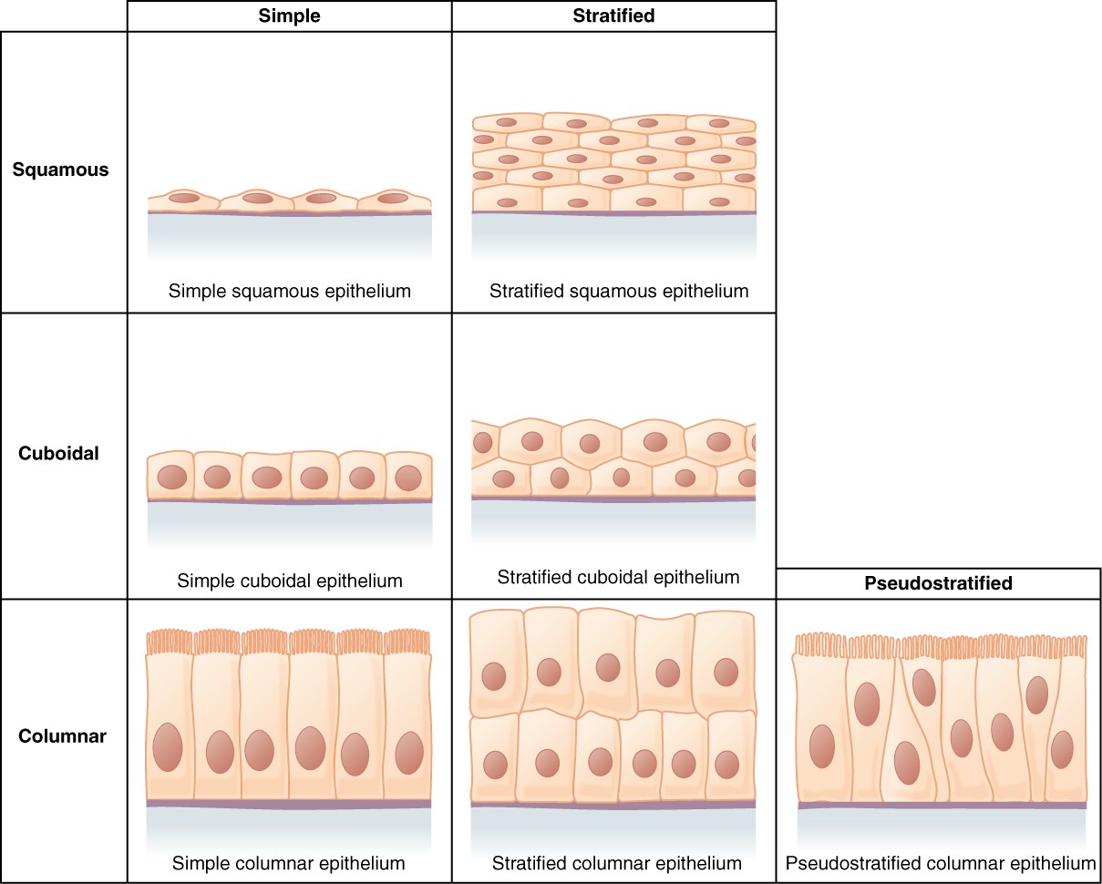
Figure 4.5 Cells of Epithelial Tissue Simple epithelial tissue is organised as a single layer of cells and stratified epithelial tissue is formed by several layers of cells.
Simple Epithelium
The shape of the cells in the single cell layer of simple epithelium reflects the functioning of those cells.
Both simple and pseudostratified columnar epithelia are heterogeneous epithelia because they include additional types of cells interspersed among the epithelial cells. For example, a goblet cell is a mucous-secreting unicellular “gland” interspersed between the columnar epithelial cells of mucous membranes ( Figure 4.6).
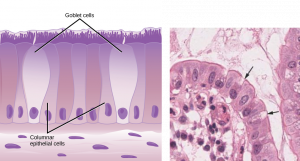
Figure 4.6 Goblet Cell (a) In the lining of the small intestine, columnar epithelium cells are interspersed with goblet cells. (b) The arrows in this micrograph point to the mucous-secreting goblet cells. LM × 1600. [credit a: Wikimedia commons, credit b:Micrograph provided by the Regents of University of Michigan Medical School © 2012]
Stratified Epithelium
A stratified epithelium consists of several stacked layers of cells. This epithelium protects against physical and chemical wear and tear. The stratified epithelium is named by the shape of the most apical layer of cells, closest to the free space. Stratified squamous epithelium is the most common type of stratified epithelium in the human body. The apical cells are squamous, whereas the basal layer contains either columnar or cuboidal cells. The top layer may be covered with dead cells filled with keratin. Mammalian skin is an example of this dry, keratinised, stratified squamous epithelium. The lining of the mouth cavity is an example of an unkeratinised, stratified squamous epithelium. Stratified cuboidal epithelium and stratified columnar epithelium can also be found in certain glands and ducts, but are uncommon in the human body.
Another kind of stratified epithelium is transitional epithelium, so-called because of the gradual changes in the shapes of the apical cells as the urinary bladder fills with urine. It is found only in the urinary system, specifically the ureters and urinary bladder. When the urinary bladder is empty, this epithelium is convoluted and has cuboidal apical cells with convex, umbrella shaped, apical surfaces. As the urinary bladder fills with urine, this epithelium loses its convolutions and the apical cells transition from cuboidal to squamous. It appears thicker and more multi-layered when the urinary bladder is empty, and more stretched out and less stratified when the urinary bladder is full and distended. Figure 4.7 summarizes the different categories of epithelial cell tissue cells.
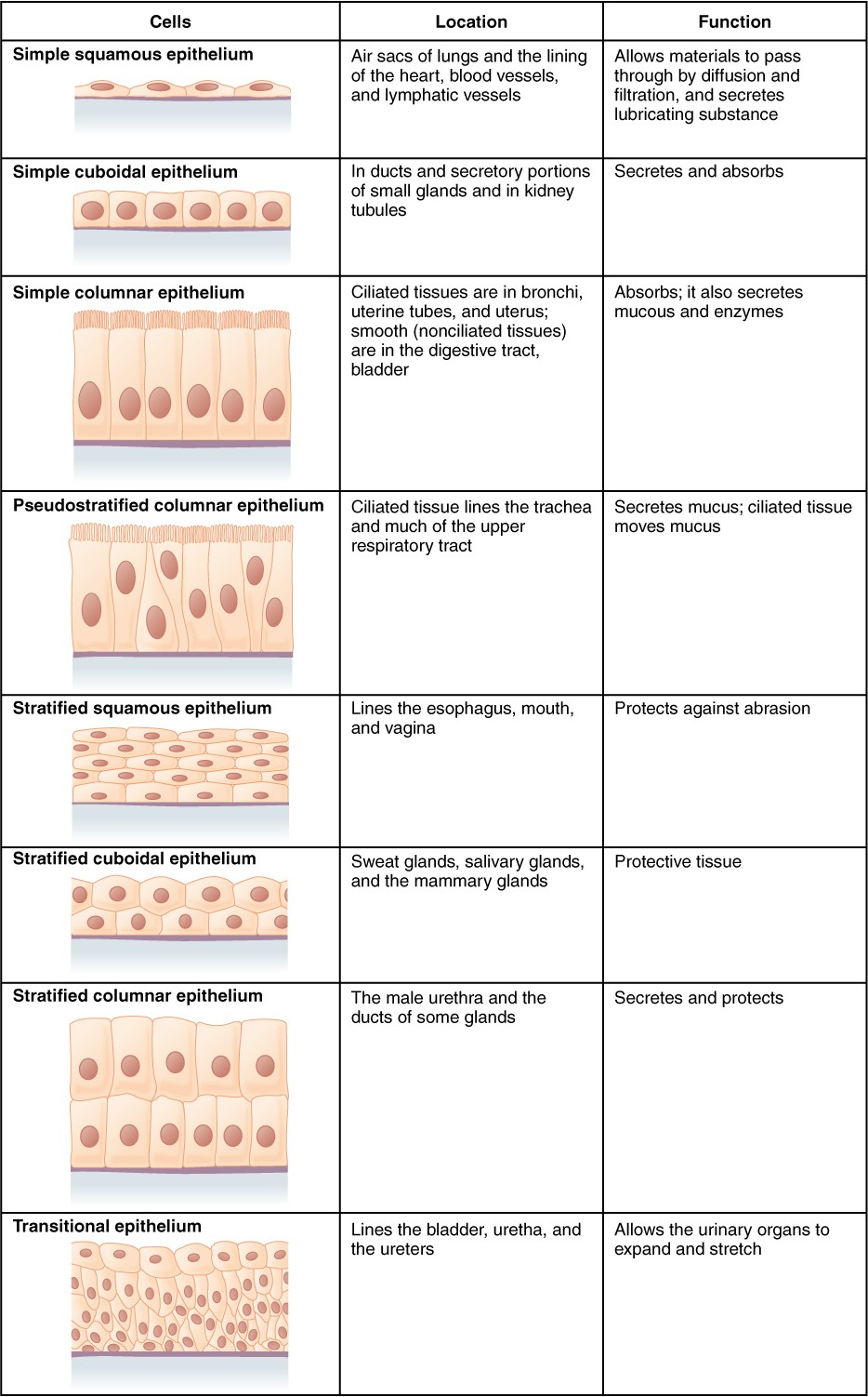
Figure 4.7 Summary of Epithelial Tissue Cells
INTERACTIVE ACTIVITY
Watch this video Review of epithelial tissues (3:50 minutes) to find out more about the anatomy of epithelial tissues.
Questions:
(a) Where in the body would you find non-keratinised stratified squamous epithelium?
INTERACTIVE ACTIVITY
Epithelial tissue classification. This activity requires you to type an answer onto the front of the Flashcard and select the Check button. The Flashcard will display immediate feedback before the next Flashcard is presented.
Questions:
(a) Consider the differing functions of the integument (skin) and the oral cavity and explain why the epidermis is comprised of keratinised stratified squamous epithelium and the oral cavity by non-keratinised stratified squamous epithelium.
(b) Why would alveoli be comprised of only simple squamous epithelium and not stratified squamous epithelium?
Glandular Epithelium
A gland is a structure made up of one or more cells modified to synthesize and secrete chemical substances. Most glands consist of groups of epithelial cells. A gland can be classified as an endocrine gland, a ductless gland that releases secretions directly into surrounding tissues and fluids (endo- = “inside”), or an exocrine gland whose secretions leave through a duct that opens directly, or indirectly, to the external environment (exo- = “outside”).
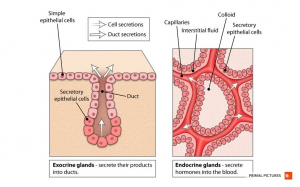
Figure 4.8 Glandular epithelium Endocrine glands secrete hormones into the blood and exocrine glands secrete chemicals at the apical surface of the epithelium. [Anatomy.TV, Primal Pictures]
Key Terms
apical that part of a cell or tissue which, in general, faces an open space
basal lamina thin extracellular layer that lies underneath epithelial cells and separates them from other tissues
basement membrane in epithelial tissue, a thin layer of fibrous material that anchors the epithelial tissue to the underlying connective tissue; made up of the basal lamina and reticular lamina
cutaneous membrane skin; epithelial tissue made up of a stratified squamous epithelial cells that cover the outside of the body
endocrine gland groups of cells that release chemical signals into the intercellular fluid to be picked up and transported to their target organs by blood
endothelium tissue that lines vessels of the lymphatic and cardiovascular system, made up of a simple squamous epithelium
epithelial membrane epithelium attached to a layer of connective tissue
epithelial tissue type of tissue that serves primarily as a covering or lining of body parts, protecting the body; it also functions in absorption, transport, and secretion
exocrine gland group of epithelial cells that secrete substances through ducts that open to the skin or to internal body surfaces that lead to the exterior of the body
goblet cell unicellular gland found in columnar epithelium that secretes mucous
ground substance fluid or semi-fluid portion of the matrix
mesothelium simple squamous epithelial tissue which covers the major body cavities and is the epithelial portion of serous membranes
mucous membrane tissue membrane that is covered by protective mucous and lines tissue exposed to the outside environment
pseudostratified columnar epithelium tissue that consists of a single layer of irregularly shaped and sized cells that give the appearance of multiple layers; found in ducts of certain glands and the upper respiratory tract
serous gland group of cells within the serous membrane that secrete a lubricating substance onto the surface
serous membrane type of tissue membrane that lines body cavities and lubricates them with serous fluid
simple columnar epithelium tissue that consists of a single layer of column-like cells; promotes secretion and absorption in tissues and organs
simple cuboidal epithelium tissue that consists of a single layer of cube-shaped cells; promotes secretion and absorption in ducts and tubules
simple squamous epithelium tissue that consists of a single layer of flat scale-like cells; promotes diffusion and filtration across surface
stratified columnar epithelium tissue that consists of two or more layers of column-like cells, contains glands and is found in some ducts
stratified cuboidal epithelium tissue that consists of two or more layers of cube-shaped cells, found in some ducts
stratified squamous epithelium tissue that consists of multiple layers of cells with the most apical being flat scale- like cells; protects surfaces from abrasion
transitional epithelium form of stratified epithelium found in the urinary tract, characterized by an apical layer of cells that change shape in response to the presence of urine
