
Chapter 14: ENDOCRINE SYSTEM
Introduction

A giant and a dwarf Abnormal levels of human growth hormone production from the anterior pituitary gland prior to puberty can lead to gigantism (excess human growth hormone production) or dwarfism (insufficient human growth hormone production. [Wellcome Images]
Chapter Objectives
After studying this chapter, you will be able to:
- Summarise the site of production of the hormones of the pituitary, thyroid, parathyroid, adrenal, pineal glands, the exocrine pancreas, and the male and female gonads
- Discuss the effects of aging on the endocrine system
Communication is a process in which a sender transmits signals to one or more receivers to control and coordinate actions. In the human body, two major organ systems participate in relatively “long distance” communication: the nervous system and the endocrine system. Together, these two systems are primarily responsible for maintaining homeostasis in the body.
The nervous system uses electrical and chemical intercellular communication. In contrast, the endocrine system uses just one method of communication: chemical signalling. These signals are sent by the endocrine organs, which secrete chemicals—the hormone—into the extracellular fluid. Hormones are transported primarily via the bloodstream throughout the body, where they bind to receptors on target cells, inducing a characteristic response. As a result, endocrine signalling requires more time than neural signalling to prompt a response in target cells, though the precise amount of time varies with different hormones.
Endocrine signalling
Learning Objectives
By the end of this section, you will be able to:
- Compare and contrast signalling in the endocrine and nervous systems
Endocrine signalling is typically less specific than neural signalling. The same hormone may play a role in a variety of different physiological processes depending on the target cells involved. For example, the hormone oxytocin promotes uterine contractions in women in labor. It is also important in breastfeeding, and may be involved in the sexual response and in feelings of emotional attachment in both males and females.
In general, the nervous system involves quick responses to rapid changes in the external environment, and the endocrine system is usually slower acting—taking care of the internal environment of the body, maintaining homeostasis, and controlling reproduction (Table 17.1). So how does the fight-or-flight response that was mentioned earlier happen so quickly if hormones are usually slower acting? It is because the two systems are connected. It is the fast action of the nervous system in response to the danger in the environment that stimulates the adrenal glands to secrete their hormones. As a result, the nervous system can cause rapid endocrine responses to keep up with sudden changes in both the external and internal environments when necessary.
Table 17.1 Endocrine and Nervous Systems
| Feature | Endocrine system | Nervous system |
| Signalling mechanism(s) | Chemical | Chemical/electrical |
| Primary chemical signal | Hormones | Neurotransmitters |
| Distance traveled | Long or short | Always short |
| Response time | Fast or slow | Always fast |
| Environment targeted | Internal | Internal and external |
Structures of the Endocrine System
Learning Objectives
By the end of this section, you will be able to:
- Describe the structures of the endocrine system
The endocrine system consists of cells, tissues, and organs that secrete hormones as a primary or secondary function. The endocrine gland is the major player in this system. The primary function of these ductless glands is to secrete their hormones directly into the surrounding fluid. The interstitial fluid and the blood vessels then transport the hormones throughout the body. The glands of the endocrine system include the pituitary, thyroid, parathyroid, adrenal (suprarenal), and pineal glands (Figure 17.2). Some of these glands have both endocrine and non-endocrine functions. The hypothalamus, pancreas, ovaries, and testes are other organs that contain cells with endocrine function.
INTERACTIVE ACTIVITY
Learn the location of endocrine organs in the brain. This activity requires you to move from one slide to the next. You can do this by clicking either the arrows beside the image, or the buttons below the image to view the unlabelled and labelled image panels of the midsagittal view of the brain. [Created in Anatomy.TV, Primal Pictures]
Question:
(a) Describe the anatomical relationship of the pituitary gland to the hypothalamus.
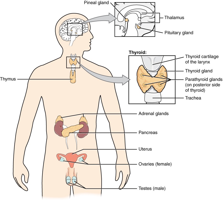
Figure 17.2 Endocrine System Endocrine glands and cells are located throughout the body and play an important role in homeostasis.
The ductless endocrine glands are not to be confused with the body’s exocrine system, whose glands release their secretions through ducts. Examples of exocrine glands include the sebaceous and sweat glands of the skin. The pancreas also has an exocrine function: most of its cells secrete pancreatic juice through the pancreatic and accessory ducts to the lumen of the small intestine.
Hormones
Learning Objectives
By the end of this section, you will be able to:
- List some common hormones produced by endocrine glands and organs
Although a given hormone may travel throughout the body in the bloodstream, it will affect the activity only of its target cells; that is, cells with receptors for that particular hormone. Once the hormone binds to the receptor, a chain of events is initiated that leads to the target cell’s response. Hormones play a critical role in the regulation of physiological processes because of the target cell responses they regulate. These responses contribute to human reproduction, growth and development of body tissues, metabolism, fluid, and electrolyte balance, sleep, and many other body functions. The major hormones of the human body and their effects are identified in Table 17.2.
Table 17.2 Endocrine Glands and Their Major Hormones
| Endocrine gland | Associated hormones | Effect |
| Pituitary (anterior) | Growth hormone (GH | Promotes growth of body tissues |
| Pituitary (anterior) | Prolactin (PRL) | Promotes milk production |
| Pituitary (anterior) | Thyroid-stimulating hormone (TSH) | Stimulates thyroid hormone release |
| Pituitary (anterior) | Adrenocorticotropic hormone (ACTH) | Stimulates hormone release by adrenal cortex |
| Pituitary (anterior) | Follicle-stimulating hormone (FSH) | Stimulates gamete production |
| Pituitary (anterior) | Luteinizing hormone (LH) | Stimulates androgen production by gonads |
| Pituitary (anterior) | Antidiuretic hormone (ADH) | Stimulates water reabsorption by kidneys |
| Pituitary (anterior) | Oxytocin | Stimulates uterine contractions during childbirth |
| Thyroid | Thyroxine (T4), triiodothyronine (T3) | Stimulate basal metabolic rate |
| Thyroid | Calcitonin | Reduces blood Ca2+ levels |
| Parathyroid | Parathyroid hormone (PTH) | Increases blood Ca2+ levels |
| Adrenal (cortex) | Aldosterone | Increases blood Na+ levels |
| Adrenal (cortex) | Cortisol, corticosterone, cortisone | Increase blood glucose levels |
| Adrenal (medulla) | Epinephrine, norepinephrine | Stimulate fight-or-flight response |
| Pineal |
Melatonin |
Regulates sleep cycles |
| Pancreas | Insulin | Reduces blood glucose levels |
| Pancreas | Glucagon | Increases blood glucose levels |
| Testes | Testosterone | Stimulates development of male secondary sex characteristics and sperm production |
| Ovaries | Oestrogens and progesterone | Stimulate development of female secondary sex characteristics and prepare the body for childbirth |
The Pituitary Gland and Hypothalamus
Learning Objectives
By the end of this section, you will be able to:
- Explain the interrelationships of the anatomy and functions of the hypothalamus and the posterior and anterior lobes of the pituitary gland
- Identify the two hormones released from the posterior pituitary
- Identify examples of hormones produced by the anterior lobe of the pituitary gland
The hypothalamus–pituitary complex can be thought of as the “command center” of the endocrine system. This complex secretes several hormones that directly produce responses in target tissues, as well as hormones that regulate the synthesis and secretion of hormones of other glands. In addition, the hypothalamus–pituitary complex coordinates the messages of the endocrine and nervous systems. In many cases, a stimulus received by the nervous system must pass through the hypothalamus–pituitary complex to be translated into hormones that can initiate a response.
The hypothalamus is a structure of the diencephalon of the brain located anterior and inferior to the thalamus (Figure 17.7). It has both neural and endocrine functions, producing and secreting many hormones. In addition, the hypothalamus is anatomically and functionally related to the pituitary gland (or hypophysis), a bean-sized organ suspended from it by a stem called the infundibulum (or pituitary stalk). The pituitary gland is cradled within the sella turcica of the sphenoid bone of the skull. It consists of two lobes that arise from distinct parts of embryonic tissue: the posterior pituitary is neural tissue, whereas the anterior pituitary is glandular tissue. The hormones secreted by the posterior and anterior pituitary are summarised in Table 17.3.
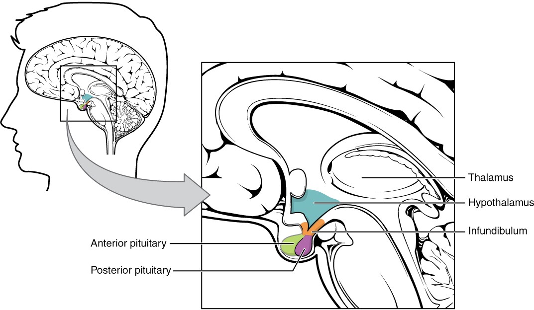
Figure 17.7 Hypothalamus–Pituitary Complex The hypothalamus region lies inferior and anterior to the thalamus. It connects to the pituitary gland by the stalk-like infundibulum. The pituitary gland consists of an anterior and posterior lobe, with each lobe secreting different hormones in response to signals from the hypothalamus.
INTERACTIVE ACTIVITY
Explore the image panels of the Pituitary Gland. This activity requires you to move from one slide to the next. You can do this by clicking either the arrows beside the image, or the buttons below the image.
The anterior portion of the pituitary gland is comprised of epithelial tissue [1st panel]. The posterior pituitary gland is comprised of nervous tissue originating in the hypothalamus [2nd panel]. [Created in Anatomy.TV, Primal Pictures]
Posterior Pituitary
The posterior pituitary is an extension of the neurons of the hypothalamus. The cell bodies of these regions rest in the hypothalamus, but their axons descend through the infundibulum, and end in the posterior pituitary (Figure 17.8). The posterior pituitary gland does not produce hormones, but rather stores and secretes the hormones antidiuretic hormone and oxytocin produced by the hypothalamus (Table …).
Anterior Pituitary
The anterior pituitary produces hormones. The anterior pituitary produces several hormones, including growth hormone (GH), thyroid-stimulating hormone (TSH), adrenocorticotropic hormone (ACTH), follicle-stimulating hormone (FSH), luteinizing hormone (LH), beta endorphin, and prolactin. (Table …).
Table 17.3 Pituitary Hormones
| Pituitary | Associated hormones | Chemical class | Effect |
| Anterior | Growth hormone (GH) | Peptide | Promotes growth of body tissues |
| Anterior | Prolactin (PRL) | Peptide | Promotes milk production from mammary glands |
| Anterior | Thyroid-stimulating hormone (TSH) | Glycoprotein | Stimulates thyroid hormone release from thyroid |
| Anterior | Adrenocorticotropic hormone (ACTH) | Peptide | Stimulates hormone release by adrenal cortex |
| Anterior | Follicle-stimulating hormone (FSH) | Glycoprotein | Stimulates gamete production in gonads |
| Anterior | Luteinizing hormone (LH) | Glycoprotein | Stimulates androgen production by gonads |
| Posterior | Antidiuretic hormone (ADH) | Peptide | Stimulates water reabsorption by kidneys |
| Posterior | Oxytocin | Peptide | Stimulates uterine contractions during childbirth |
The Thyroid Gland
Learning Objectives
By the end of this section, you will be able to:
- Describe the location and anatomy of the thyroid gland
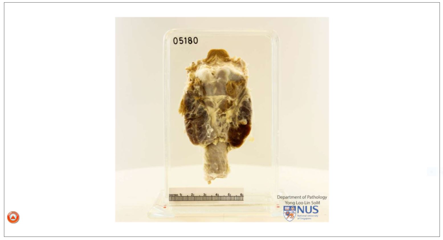
A butterfly-shaped organ, the thyroid gland is located anterior to the trachea, just inferior to the larynx (Figure 17.12). The medial region, called the isthmus, is flanked by wing-shaped left and right lobes. Each of the thyroid lobes are embedded with parathyroid glands, primarily on their posterior surfaces. The thyroid gland produces thyroid hormones and calcitonin (Table ..).
INTERACTIVE ACTIVITY
Learn the location of Thyroid gland. This activity requires you to move from one slide to the next. You can do this by clicking either the arrows beside the image, or the buttons below the image to view the unlabelled and labelled image panels of the trachea, tracheal cartilages and thyroid gland. [Created in Anatomy.TV, Primal Pictures]
INTERACTIVE LINK
The Parathyroid Glands
Learning Objectives
By the end of this section, you will be able to:
- Describe the location and structure of the parathyroid glands
The parathyroid glands are tiny, round structures usually found embedded in the posterior surface of the thyroid gland (Figure 17.15). A thick connective tissue capsule separates the glands from the thyroid tissue. Most people have four parathyroid glands, but occasionally there are more in tissues of the neck or thorax. These glands produce and secrete parathyroid hormone (PTH), the major hormone involved in the regulation of blood calcium levels. (Table …).
INTERACTIVE ACTIVITY
The Adrenal (Suprarenal) Glands
Learning Objectives
By the end of this section, you will be able to:
- Describe the location and structure of the adrenal glands
The adrenal (suprarenal) glands are wedges of glandular and neuroendocrine tissue attached to the superior surface of the kidneys by a fibrous capsule (Figure 17.17). The adrenal gland consists of an outer cortex of glandular tissue and an inner medulla of nervous tissue. Each region secretes its own set of hormones. These glands produce and secrete epinephrine (adrenaline) and norepinephrine (noradrenaline) which stimulate the fight or flight response. (Table …).
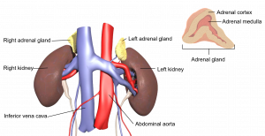
Figure 17.17 Adrenal Glands Both adrenal glands are located superior to the kidneys and are composed of an outer cortex and an inner medulla, all surrounded by a connective tissue capsule. [WikiMedia Commons]
The Pineal Gland
Learning Objectives
By the end of this section, you will be able to:
- Describe the location and structure of the pineal gland
Recall that the hypothalamus, part of the diencephalon of the brain, is located inferior and anterior to the thalamus. Inferior and posterior to the thalamus is the pineal gland, a tiny endocrine gland whose functions are not entirely clear. The pineal gland produces and secretes the hormone melatonin to regulate sleep cycles. (Table …).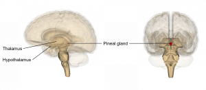
Figure 17.17 The pineal gland The pineal gland is located inferior and posterior to the thalamus. [Life Science Databases]
The Pancreas
Learning Objectives
By the end of this section, you will be able to:
- Describe the location and structure of the pancreas, and the morphology and function of the pancreatic islets
The pancreas is a long, slender organ located posterior to the stomach (Figure 17.18). Although it is primarily an exocrine gland, secreting a variety of digestive enzymes, the pancreas has an endocrine function. Its pancreatic islets—clusters of cells formerly known as the islets of Langerhans—secrete the hormones glucagon and insulin which together regulate blood glucose levels.
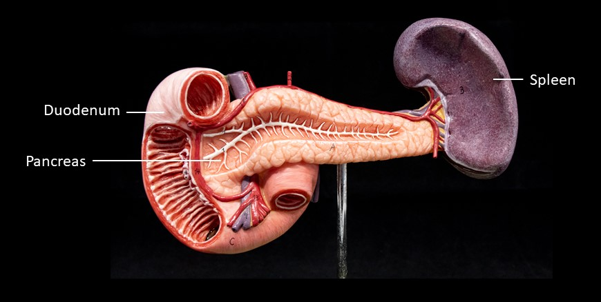
Figure 17.18 Pancreas The pancreas extends form the duodenum to the spleen.
The Male and Female Gonads
Learning Objectives
By the end of this section, you will be able to:
- Identify the gonadal organs of the endocrine system and their location in the body
INTERACTIVE ACTIVITY
Key Terms
adrenal cortex outer region of the adrenal glands consisting of multiple layers of epithelial cells and capillary networks
adrenal glands endocrine glands located superior to each kidney that are important for the regulation of the stress response, blood pressure and blood volume, water homeostasis, and electrolyte levels
adrenal medulla inner layer of the adrenal glands that plays an important role in the stress response by producing epinephrine and norepinephrine
aldosterone hormone produced and secreted by the adrenal cortex that stimulates sodium and fluid retention and increases blood volume and blood pressure
antidiuretic hormone (ADH) hypothalamic hormone that is stored by the posterior pituitary and that signals the kidneys to reabsorb water
calcitonin peptide hormone produced and secreted by the thyroid gland that functions to decrease blood calcium levels
endocrine gland tissue or organ that secretes hormones into the blood and lymph without ducts such that they may be transported to organs distant from the site of secretion
endocrine system cells, tissues, and organs that secrete hormones as a primary or secondary function and play an integral role in normal bodily processes
epinephrine hormone secreted by the adrenal medulla in response to short- term stress; also called adrenaline
oestrogens class of predominantly female sex hormones important for the development and growth of the female reproductive tract, secondary sex characteristics, the female reproductive cycle, and the maintenance of pregnancy
exocrine system cells, tissues, and organs that secrete substances directly to target tissues via glandular ducts
follicle-stimulating hormone (FSH) anterior pituitary hormone that stimulates the production and maturation of sex cells
glucagon pancreatic hormone that stimulates the catabolism of glycogen to glucose, thereby increasing blood glucose levels
growth hormone (GH) anterior pituitary hormone that promotes tissue building and influences nutrient metabolism
hormone secretion of an endocrine organ that travels via the bloodstream or lymphatics to induce a response in target cells or tissues in another part of the body
hormone receptor protein within a cell or on the cell membrane that binds a hormone, initiating the target cell response
hypothalamus region of the diencephalon inferior to the thalamus that functions in neural and endocrine signalling
infundibulum stalk containing vasculature and neural tissue that connects the pituitary gland to the hypothalamus (also called the pituitary stalk)
insulin pancreatic hormone that enhances the cellular uptake and utilisation of glucose, thereby decreasing blood glucose levels
luteinizing hormone (LH) anterior pituitary hormone that triggers ovulation and the production of ovarian hormone in females, and the production of testosterone in males
melatonin hormone that is secreted in response to low light and causes drowsiness
norepinephrine hormone secreted by the adrenal medulla in response to short-term stress; also called noradrenaline
oxytocin hypothalamic hormone stored in the posterior pituitary gland and important in stimulating uterine contractions in labor, milk ejection during breastfeeding, and feelings of attachment (also produced in males)
pancreas organ with both exocrine and endocrine functions located posterior to the stomach that is important for digestion and the regulation of blood glucose
pancreatic islets specialised clusters of pancreatic cells that have endocrine functions; also called islets of Langerhans
parathyroid glands small, round glands embedded in the posterior thyroid gland that produce parathyroid hormone (PTH)
parathyroid hormone (PTH) blood calcium levels hormone produced and secreted by the parathyroid glands in response to low blood calcium levels
pineal gland endocrine gland that secretes melatonin, which is important in regulating the sleep-wake cycle
pituitary gland bean-sized organ suspended from the hypothalamus that produces, stores, and secretes hormones in response to hypothalamic stimulation (also called hypophysis)
progesterone predominantly female sex hormone important in regulating the female reproductive cycle and the maintenance of pregnancy
prolactin (PRL) anterior pituitary hormone that promotes development of the mammary glands and the production of breast milk
testosterone hormone secreted by the male testes and important in the maturation of sperm cells, growth and development of the male reproductive system, and the development of male secondary sex characteristics
thyroid gland large endocrine gland responsible for the synthesis of thyroid hormones