
Chapter 9: MUSCULAR SYSTEM
Introduction
A Body in Motion The muscular system allows us to move, flex and manipulate our bodies in space. Think about the things that you do each day—talking, walking, sitting, standing, and running—all of these activities require movement of particular skeletal muscles. [GoodFon.com]
Chapter Objectives
After studying this chapter, you will be able to:
- Describe the actions and roles of agonists and antagonists
- Explain the structure and organisation of muscle fascicles and their role in generating force
- Explain the criteria used to name skeletal muscles
- Identify the skeletal muscles and their actions on the skeleton and soft tissues of the body
- Identify the origins and insertions of skeletal muscles and their prime movements
There are approximately six hundred skeletal muscles in the human body which account for forty percent of the total body weight. Skeletal muscles are organs that contain the four types of primary tissues. The major component is skeletal muscle fibres that are contained within the muscle belly. Dense irregular conenctive tissue surrounds each skeletal muscle as deep fascia and dense regular connective tissue is contained within its tendons and/or aponeuroses. Areolar connective tissue, blood vessels and their epithelial lining, nerves, nerve receptors and nerve endings make up the minor components.
The system to name skeletal muscles is known as muscle nomenclature; in some cases, the muscle is named by its shape, and in other cases it is named by its location or attachments to the skeleton. If you understand the meaning of the name of the muscle, often it will help you remember its location and/or what it does. This chapter also will describe how skeletal muscles are arranged to accomplish movement, and how other muscles may assist, or be arranged on the skeleton to resist or carry out the opposite movement. The actions of the skeletal muscles will be covered in a regional manner, working from the head down to the toes.
Skeletal Muscle Organisation
By the end of this section, you will be able to:
- Define the terms origin and insertion in terms of how they apply to the attachments of skeletal muscles
- Describe the role of an agonist and an antagonist
The connective tissue component of skeletal muscles plays a very important role in the formation of muscle attachments. Each skeletal muscle fibre, each bundle of muscle fibres and each muscle is surrounded by specific connective tissue sheaths that help to organise the muscle organ and allow passageways for nerves and blood vessels to reach the muscle fibres. Deep fascia is a dense irregular connective tissue sheath that surrounds groups of individual muscle that share a common function and serves to compartmentalise muscles into functional groups. A skeletal muscle compartment shares a common action/ joint movement, innervation and blood supply.
Each muscle belly attaches to the skeleton and/or adjacent tissues by a tendon, aponeurosis or fleshy attachment.
To pull on a bone, that is, to change the angle at its synovial joint, which essentially moves the skeleton, a skeletal muscle must also be attached to a fixed part of the skeleton. The moveable end of the muscle that attaches to the bone being pulled is called the muscle’s insertion, and the end of the muscle attached to a fixed (stabilised) bone is called the origin. For example, as an elbow flexor, the brachialis has its origin on the humeral shaft (fixed point that does not move during joint movement) and its insertion on the ulna (contraction of brachialis moves ulna anteriorly towards humerus to flex elbow; bony attachment that moves).
There are also skeletal muscles that do not pull against the skeleton for movements. For example, there are the muscles that produce facial expressions. The insertions and origins of facial muscles are in the skin, so that certain individual muscles contract to form a smile or frown, form sounds or words, and raise the eyebrows. There also are skeletal muscles in the tongue, and the external urinary and anal sphincters that allow for voluntary regulation of urination and defecation, respectively. In addition, the thoracic diaphragm contracts and relaxes to change the volume of the pleural cavities but it does not move the skeleton to do this.
Although a number of muscles may be involved in any one joint movement, the principal muscle involved is called the prime mover, or agonist. A muscle with the opposite action of the prime mover is called an antagonist. Antagonists play two important roles in muscle function: (1) they maintain body or limb position, such as holding the upper limb straight (elbow extended) or standing erect; and (2) they control rapid movement, to support the coordinated motion of a limb. For example, to extend the knee joint, a group of four muscles called the quadriceps femoris in the anterior compartment of the thigh are activated (and would be called the agonists of knee extension). However, to flex the knee joint, an opposite or antagonistic set of muscles called the hamstrings is activated. See Table 24.1 for a list of some agonists and antagonists.
Table 24.1 Agonist and Antagonist Skeletal Muscle Pairs
| Agonist | Antagonist | Movement |
| Biceps brachii: in the anterior compartment of the brachium | MovementBiceps brachii: in the anterior compartment of the brachiumTriceps brachii: in the posterior compartment of the brachium | The biceps brachii flexes the antebrachium, whereas the triceps brachii extends it. |
| Hamstrings: group of three muscles in the posterior compartment of the thigh | Quadriceps femoris: group of four muscles in the anterior compartment of the thigh | The hamstrings flex the leg, whereas the quadriceps femoris extend it. |
| Flexor digitorum superficialis and flexor digitorum profundus: in the anterior compartment of the antebrachium | Extensor digitorum: in the posterior compartment of the antebrachium | The flexor digitorum superficialis and flexor digitorum profundus flex the fingers and the hand at the wrist, whereas the extensor digitorum extends the fingers and the hand at the wrist. |
Skeletal Muscle Nomenclature
By the end of this section, you will be able to:
- Describe the criteria used to name skeletal muscles
- Explain how understanding the muscle names helps describe shapes, location, and actions of various muscles
Anatomists name the skeletal muscles according to a number of criteria, each of which describes the muscle in some way. These include naming the muscle after its shape, its size compared to other muscles in the area, its location in the body or the location of its attachments to the skeleton, how many origins it has, or its action. When you understand the names of muscles it will help you remember where the muscles are located and what they do (Figure 24.6). Most muscles are named according to a combination of these criteria.
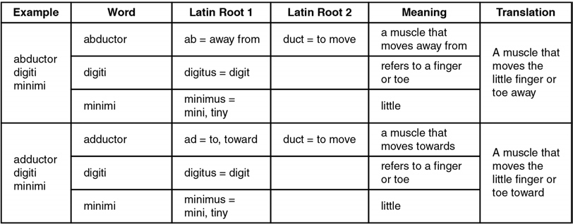
Figure 24.6 Understanding a Muscle Name from the Latin
Axial Muscles
Learning Objectives
By the end of this section, you will be able to:
- Identify examples of axial muscles of the face, head and neck
- Identify examples of axial muscles of the thorax and abdomen
- Describe the features of axial skeletal muscles from their nomenclature
The skeletal muscles are divided into axial (muscles of the trunk and head) and appendicular (muscles of the upper and lower limbs) categories. This system reflects the bones of the skeletal system, which are also arranged in this manner.
Skeletal Muscles of the Head and Neck
The origins of the muscles of facial expression are on the surface of the skull (remember, the origin of a muscle does not move). The insertions of these muscles have fibres intertwined with connective tissue and the dermis of the skin. Because the muscles insert in the skin rather than on bone, when they contract, the skin moves to create facial expression (Figure 24.7). Muscles of facial expression include the orbicularis oris, a circular muscle that moves the lips, and the orbicularis oculi, a circular muscle that closes the eye.
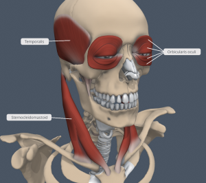
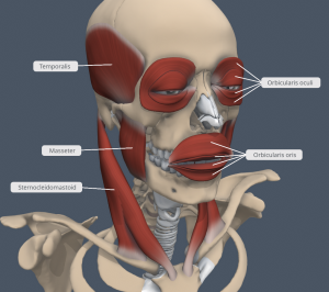
Figure 24.7 Muscles of Facial Expression Many of the muscles of facial expression insert into the skin surrounding the eyelids and mouth, producing facial expressions by moving the skin rather than bones. The muscles of mastication assist in the movement of the temporomandibular joint. The sternocleidomastoid muscle attaches between the sternum, clavicle and mastoid process of the temporal bone. [Created in Anatomy.TV, Primal Pictures]
In anatomical terminology, chewing is called mastication. Muscles involved in chewing must be able to exert enough pressure to bite through and then chew food before it is swallowed (Figure 24.10 and Table 24.4). Muscles involved in chewing must be able to exert enough pressure to bite through and then chew food before it is swallowed (Figure 24.10 and Table 24.4). The masseter muscle is the main muscle used for chewing because it elevates the mandible at the temporomandibular joint (lower jaw) to close the mouth, and it is assisted by the temporalis muscle, which retracts the mandible. You can feel the temporalis move by putting your fingers to your temple as you chew.
The head, attached superior to the vertebral column, is balanced, moved, and rotated by the neck muscles (Table 24.5). When these muscles act unilaterally, the head rotates. When they contract bilaterally, the head flexes or extends. The major muscle that laterally flexes and rotates the head is the sternocleidomastoid. In addition, both muscles working together are the flexors of the head. Place your fingers on both sides of the neck and turn your head to the left and to the right. You will feel the movement originate there.
Skeletal Muscles of the Back
The back muscles stabilise and move the vertebral column, and are grouped according to the lengths and direction of the fascicles. The trapezius is a large triangular-shaped muscle (trapezoid-shaped when right and left trapezius are observed together) located in the posterior neck and back. The broad, triangular latissimus dorsi muscle is located on the inferior part of the back, where it inserts into a thick connective tissue sheath called an aponeurosis.
The erector spinae group lies deep to the trapezius and latissimus dorsi and forms the majority of the muscle mass of the back and it is the primary extensor of the vertebral column. It controls flexion, lateral flexion, and rotation of the vertebral column, and maintains the lumbar curvature.
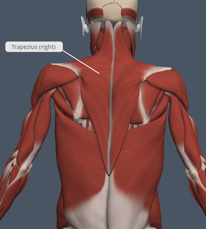
Figure 24.15 Muscles of the Back The large, complex muscles of the back move the neck, shoulders, and vertebral column. Note the diamond-shaped trapezius lying superficial on the back. [Created in Anatomy.TV, Primal Pictures]
INTERACTIVE ACTIVITY
Explore the image panels of the superficial (1st panel) and deep (2nd panel) Muscles of the Back by clicking the arrows beside each image, or the buttons below the image panel. [Created in Anatomy.TV, Primal Pictures]
Skeletal Muscles of the Thorax
The muscles of the thorax serve to facilitate breathing by changing the size of the thoracic cavity (Table 24.7). When you inhale, your chest rises because the cavity expands. Alternately, when you exhale, your chest falls because the thoracic cavity decreases in size. The change in volume of the thoracic cavity during breathing is due to the alternate contraction and relaxation of the thoracic diaphragm (Figure 24.17). It separates the thoracic and abdominal cavities, and is dome-shaped at rest. The superior surface of the thoracic diaphragm is convex, creating the elevated floor of the thoracic cavity. The inferior surface is concave, creating the curved roof of the abdominal cavity. The inferior surface of the pericardial sac and the inferior surfaces of the pleural membranes (parietal pleura) fuse onto the central tendon of the thoracic diaphragm. To the sides of the tendon are the skeletal muscle portions of the thoracic diaphragm, which insert into the tendon while having a number of origins including the xiphoid process of the sternum anteriorly, the inferior six ribs and their cartilages laterally, and the lumbar vertebrae and 12th ribs posteriorly. The thoracic diaphragm also includes three openings for the passage of structures between the thorax and the abdomen. The inferior vena cava passes through the caval foramen, and the oesophagus and attached nerves pass through the oesophageal hiatus. The descending aorta, thoracic duct, and azygos vein pass through the aortic hiatus of the posterior to the thoracic diaphragm.
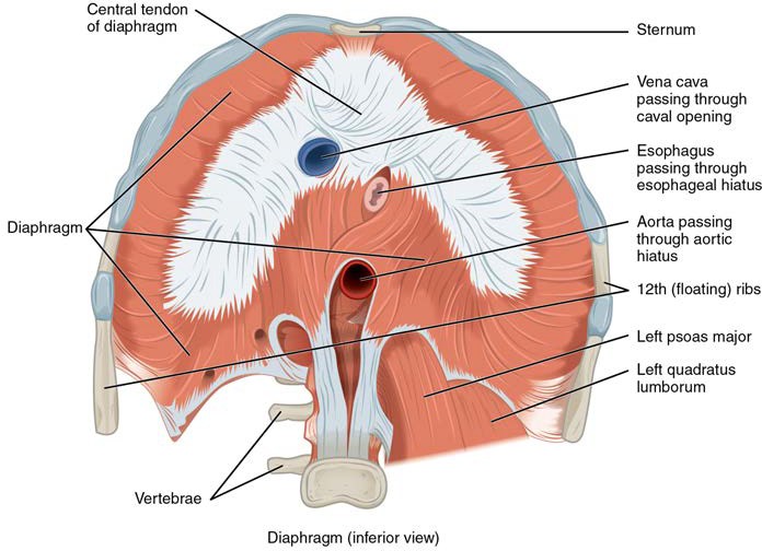
Figure 24.17 Muscles of the Thoracic Diaphragm The thoracic diaphragm separates the thoracic and abdominal cavities.
A muscle located in the thorax but associated with the movement of the glenohumeral joint is the pectoralis major. It is a thick and fan-shaped muscle, covering much of the superficial portion of the anterior thorax. The pectoralis major has two heads: a larger sternal head and a smaller more superior clavicular head. The serratus anterior muscle consists of several long slips of muscle that extend from the medial border of the scapula to attach to the ribs, giving it a serrated edge anteriorly. The muscle assists with respiration.
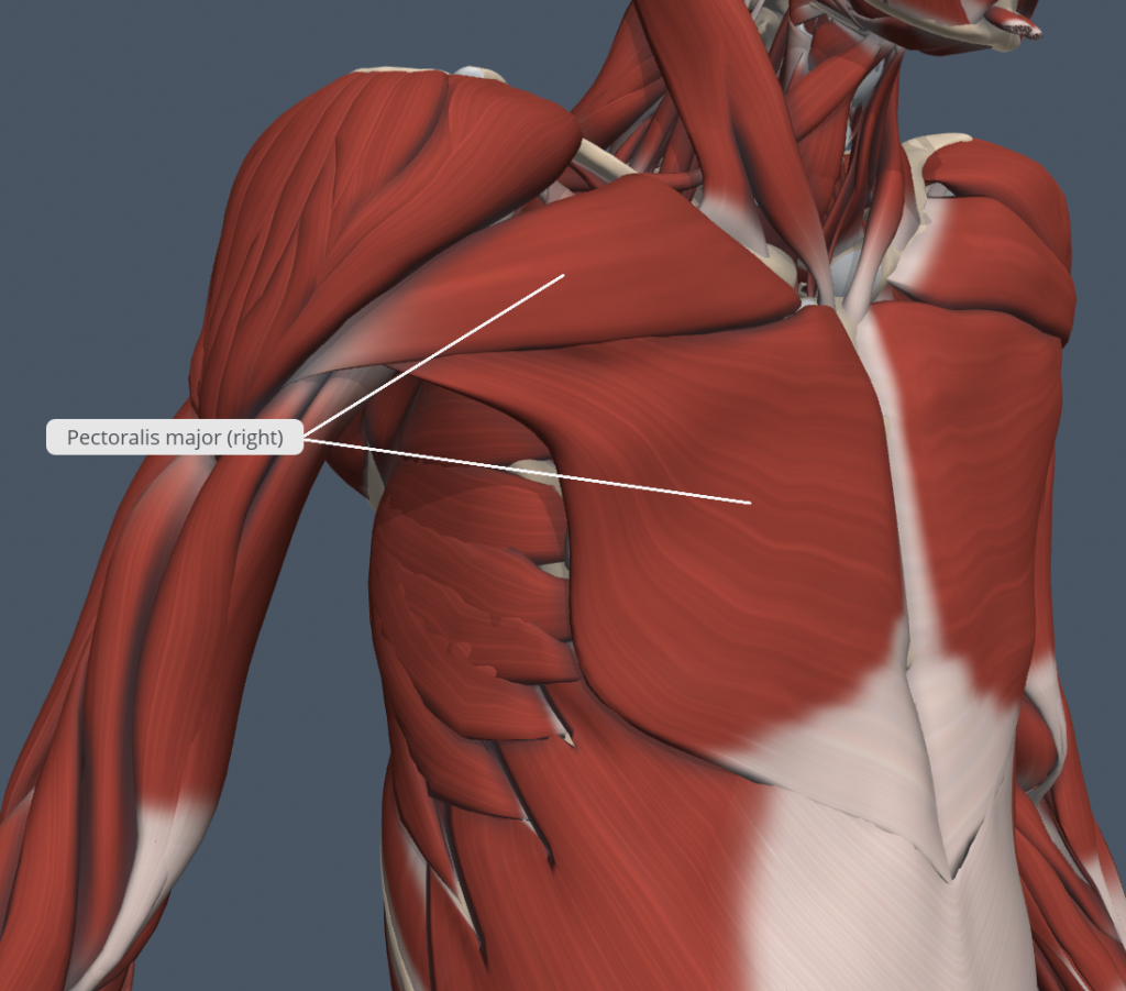
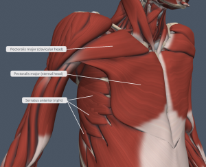
Figure 24.15 Muscles of the Anterior Thorax The large pectoralis major contributes to the movement of the glenohumeral joint whilst the serratus anterior assists with respiration. [Created in Anatomy.TV, Primal Pictures]
Skeletal Muscles of the Abdomen
There are four pairs of abdominal muscles that cover the anterior and lateral abdominal region and meet at the anterior midline. These muscles of the anterolateral abdominal wall can be divided into four muscles: the external obliquus abdominis, the internal obliquus abdominis, the transversus abdominis, and the rectus abdominis (Figure 24.16 and Table 24.6). Three of these muscles are located in the anterolateral wall of the abdomen. The external obliquus abdominis (most superficial), extends inferiorly and medially, in the direction of sliding one’s four fingers into pant pockets. Perpendicular to it is the intermediate internal obliquus abdominis, extending superiorly and medially, the direction the thumbs usually point when the other fingers are in your pant pocket. The deepest muscle, the transversus abdominis, is arranged transversely around the abdomen, similar to the front of a belt on a pair of pants. This arrangement of three bands of muscles in different orientations allows various movements and rotations of the trunk. The three layers of muscle also help to protect the internal abdominal organs in an area where there is no bone.
The rectus abdominis muscles (a pair of long, linear muscles) lie medial to the external and internal obliquus abdominis muscles and originate at the pubic symphysis, and extend the length of the body’s abdomen. Each muscle is segmented by three transverse bands of collagen fibres called the tendinous intersections. This results in the look of “six-pack abs,” as each segment hypertrophies.
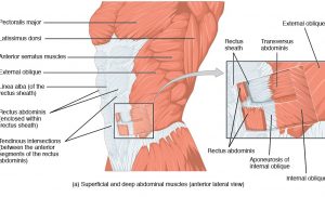
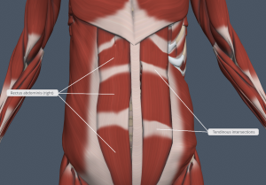
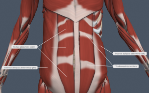
Figure 24.16 Muscles of the Abdomen The anterior abdominal muscles include the medially located rectus abdominis. Lateral to the rectus abdominis, the abdominal wall is composed of three layers. The external obliquus abdominis form the superficial layer, while the internal obliquus abdominis form the middle layer, and the transversus abdominis forms the deepest layer. [Created in Anatomy.TV, Primal Pictures]
Appendicular Muscles
Learning Objectives
By the end of this section, you will be able to:
- Identify the muscles of the pectoral girdle and upper limbs
- Identify the muscles of the lower limb
- Describe the features of appendicular skeletal muscles from their nomenclature
Skeletal Muscles of the Pectoral Girdle and Brachium (Arm)
The deltoid, the thick muscle that creates the rounded lines of the shoulder is the major abductor of the glenohumeral joint, but it also facilitates flexion and medial rotation, as well as extension and lateral rotation. The deltoid inserts onto the deltoid tuberosity of the lateral aspect of the shaft of the humerus. The subscapularis originates on the anterior scapula and medially rotates the glenohumeral joint. Named for their locations, the supraspinatus (superior to the spine of the scapula) and the infraspinatus (inferior to the spine of the scapula) abduct the glenohumeral joint, and laterally rotate the glenohumeral joint, respectively. The thick and flat teres major is inferior to the teres minor and extends the glenohumeral joint, and assists in adduction and medial rotation of it. The long teres minor laterally rotates and extends the glenohumeral. Finally, the coracobrachialis flexes and adducts the glenohumeral joint.
The tendons of the subscapularis, supraspinatus, infraspinatus, and teres minor connect the scapula to the humerus, forming the rotator cuff (musculotendinous cuff), the circle of tendons around the glenohumeral joint.
INTERACTIVE ACTIVITY
INTERACTIVE ACTIVITY
INTERACTIVE ACTIVITY
The muscles that flex the elbow include the biceps brachii, brachialis and brachioradialis. The two-headed biceps brachii crosses the glenohumeral and elbow joints to flex the elbow, supinate the radioulnar joints, whilst also taking part in flexing the glenohumeral joint. The biceps brachii consists of a more lateral ‘long head’ which has its origin on the superior part of the glenoid fossa of the scapula and a more medial ‘short head’ which has its origin on the coracoid process of the scapula. Both heads of the biceps brachii then combine into a shared tendon to insert onto the radial tuberosity.
Deep to the biceps brachii, the brachialis provides additional power in flexing the elbow joint. Finally, the brachioradialis can flex the elbow joint quickly or help lift a load slowly. These muscles and their associated blood vessels and nerves form the anterior compartment of the brachium (anterior flexor compartment of the arm) (Figure 24.25 and Figure 24.26).
The posterior compartment of the brachium are the extensors of the elbow joint and is made up of the three heads of the triceps brachii. The triceps brachii consists of a ‘long head’ that has its origin on the inferior aspect of the glenoid fossa of the scapula, and a ‘lateral head’ and ‘medial head’ that both originate from the shaft of the humerus. The three heads of the triceps brachii share a common tendon that inserts onto the olecranon of the ulna.
Skeletal Muscles of the Antebrachium (Forearm) and Hand
Radiocarpal and digit movements are facilitated by two groups of muscles. The antebrachium is the origin of the extrinsic muscles of the hand. The palm is the origin of the intrinsic muscles of the hand.
The muscles in the anterior compartment of the antebrachium (anterior flexor compartment of the antebrachium) originate on the humerus and insert onto different parts of the hand. These make up the bulk of the antebrachium. From lateral to medial, the superficial anterior compartment of the antebrachium arises from a common origin on the medial epicondyle of the humerus and includes the pronator teres, flexor carpi radialis, palmaris longus (not found in all people), flexor carpi ulnaris, and flexor digitorum superficialis. The flexor digitorum superficialis flexes the radiocarpal joint as well as the digits at the metacarpal and interphalangeal joints, which allows for rapid finger movements, as in typing or playing a musical instrument (see Figure 24.27 and Table 24.9). The pronator teres is an important pronator of the radioulnar joints. The deep anterior compartment produces flexion to move the fingers to make a fist. These include flexor pollicis longus and the flexor digitorum profundus.
The muscles in the superficial posterior compartment of the antebrachium (superficial posterior extensor compartment of the antebrachium) originate on the lateral epicondyle of the humerus. These are the extensor radialis longus, extensor carpi radialis brevis, extensor digitorum, extensor digiti minimi, and the extensor carpi ulnaris (see Figure 24.27). Also note the brachioradialis muscle which is anterolaterally located in the antebrachium. It is located in the posterior compartment of the antebrachium but is a powerful flexor of the elbow joint.
INTERACTIVE ACTIVITY
INTERACTIVE ACTIVITY
Skeletal Muscles of the Gluteal and Femoral (Thigh) Regions
The appendicular muscles of the inferior part of the body stabilise the pelvic girdle, which serves as a foundation for the lower limbs. Comparatively, there is much more movement at the pectoral girdle than at the pelvic girdle.
Most muscles that insert on the femur and move it, originate on the pelvic girdle. Some of the largest and most powerful muscles in the body are the gluteal muscles or gluteal group. The gluteus maximus is the largest of these muscles. Deep to the gluteus maximus, a triangular or pyramidal muscle is visible called the piriformis. The piriformis inserts onto the greater trochanter of the femur and forms the roof of the greater sciatic foramen through which the sciatic nerve travels to enter the posterior thigh.
The skeletal muscles of the femoral region are separated into three compartments by deep fascia: anterior (quadriceps femoris), posterior (hamstrings) and the medial adductor compartment (see Figure 24.29 and Figure 24.31). The muscles of the medial compartment of the thigh are made up of many muscles including the adductor longus and the adductor magnus. These muscles can both medially and laterally rotate the thigh depending on the placement of the foot. The adductor longus flexes the thigh, whereas the adductor magnus extends it.
The muscles of the anterior compartment of the thigh flex the hip joint and extend the knee joint. This compartment contains the quadriceps femoris group, which actually comprises four muscles that extend and stabilise the knee. The rectus femoris is on the anterior aspect of the thigh, the vastus lateralis is on the lateral aspect of the thigh, the vastus medialis is on the medial aspect of the thigh, and the vastus intermedius is between the vastus lateralis and vastus medialis and deep to the rectus femoris. The tendon common to all four is the quadriceps tendon, which inserts into the patella and continues inferior to the patella as the patellar ligament. The patellar ligament attaches to the tibial tuberosity. In addition to the quadriceps femoris, the sartorius is a band-like muscle that extends from the anterior superior iliac spine of the os coxa to the medial side of the proximal tibia. This versatile muscle flexes the leg at the knee and flexes, abducts, and laterally rotates the leg at the hip. This muscle allows us to sit cross-legged.
The posterior compartment of the thigh includes muscles that flex the knee and extend the hip joint. The three long muscles on the back of the knee are the hamstring group, which flexes the knee. These are the biceps femoris, semitendinosus, and semimembranosus. The tendons of these muscles form the boundaries of the popliteal fossa, the diamond-shaped space on the posterior surface of the knee. The hamstring muscles have a common origin on the ischial tuberosity of the os coxa.
INTERACTIVE ACTIVITY
INTERACTIVE ACTIVITY
Skeletal Muscles of the Crural (Leg) Region and Foot
Similar to the thigh muscles, the muscles of the leg are divided by deep fascia into three compartments: anterior, lateral, and posterior (Figure 24.32 and Figure 24.33).
The muscles in the anterior compartment of the leg include the tibialis anterior, a long and thick muscle on the lateral surface of the tibia. The lateral compartment of the leg includes two muscles, including the fibularis longus (peroneus longus). The superficial muscles in the posterior compartment of the leg all insert onto the calcaneal tendon (Achilles tendon), a strong tendon that inserts into the calcaneus of the tarsus. The muscles in this compartment are colloquially referred to as the calf muscles and are large and strong to keep humans upright. Due to their common insertion these muscles are collectively referred to as the triceps surae (three headed muscle of the posterior leg). The most superficial and visible muscle of the triceps surae is the gastrocnemius, which has a medial and a lateral head. Deep to the gastrocnemius is the wide, flat soleus.
The foot also has intrinsic muscles, which originate and insert within it. These muscles primarily provide support for the foot and its arch, and contribute to movements of the toes (Figure 24.34 and Figure 24.35). The intrinsic muscles of the foot consist of two groups: dorsal and plantar. The plantar group consists of many small muscles including the abductor hallucis located on the medial side of the foot and inserts onto the hallux.
INTERACTIVE ACTIVITY
Explore the image panels of the Muscles of the Leg and Foot that move the talocrural joint and digits of the foot by clicking the arrows beside each image, or the buttons below the image panel. The muscles of the crural region are organised into three compartments: anterior, lateral and posterior. Review the anterolateral (1st panel), superficial posterior (2nd panel) and deep posterior (3rd panel) views of the leg; and a plantar view of the foot (4th panel). [Created in Anatomy.TV, Primal Pictures]
Questions:
(a) Write a three column list of all of the muscles in these images belonging to the anterior, lateral and posterior compartments of the crural region.
(b) What is meant by the terms intrinsic and extrinsic muscles of the foot?
Key Terms
abduct move away from midline in the sagittal plane
abductor moves the bone away from the midline
adductor moves the bone toward the midline
adductor longus muscle that adducts, medially rotates, and flexes the thigh
adductor magnus muscle with an anterior fascicle that adducts, medially rotates and flexes the thigh, and a posterior fascicle that assists in thigh extension
agonist (also, prime mover) muscle whose contraction is responsible for producing a particular motion
antagonist muscle that opposes the action of an agonist
anterior compartment of the brachium (anterior flexor compartment of the arm) the biceps brachii, brachialis, brachioradialis, and their associated blood vessels and nerves
anterior compartment of the antebrachium (anterior flexor compartment of the forearm) deep and superficial muscles that originate on the humerus and insert into the hand
anterior compartment of the leg (crural region) region that includes muscles that dorsiflex the talocrural joint
anterior compartment of the thigh (femoral region) region that includes muscles that flex the hip joint and extend the knee joint
appendicular of the arms and legs
axial of the trunk and head
belly bulky central body of a muscle
bi two
biceps brachii two-headed muscle that crosses the glenohumeral and elbow joints to flex the elbow while assisting in supinating the radioulnar joints and flexing the glenohumeral joint
biceps femoris hamstring muscle
brachialis muscle deep to the biceps brachii that provides power in flexing the forearm.
brachioradialis muscle that can flex the forearm quickly or help lift a load slowly
brevis short
calcaneal tendon (also, Achilles tendon) strong tendon that inserts into the calcaneal bone of the ankle
caval foramen opening in the diaphragm that allows the inferior vena cava to pass through; foramen for the vena cava
coracobrachialis muscle that flexes and adducts the arm
deep anterior compartment of antebrachium flexor pollicis longus, flexor digitorum profundus, and their associated blood vessels and nerves
deep posterior compartment of antebrachium (deep posterior extensor compartment of the forearm) the abductor pollicis longus, extensor pollicis brevis, extensor pollicis longus, extensor indicis, and their associated blood vessels and nerves
deltoid shoulder muscle that abducts the arm as well as flexes and medially rotates it, and extends and laterally rotates it
dorsal muscles of foot region that includes the extensor digitorum brevis
erector spinae group large muscle mass of the back; primary extensor of the vertebral column
extensor muscle that increases the angle at the joint
extensor carpi ulnaris muscle that extends and adducts the hand
extensor digiti minimi muscle that extends the little finger
extensor digitorum muscle that extends the hand at the wrist and the phalanges
extensor digitorum brevis muscle that extends the toes
extensor digitorum longus muscle that is lateral to the tibialis anterior
extensor radialis longus muscle that extends and abducts the hand at the wrist
external obliquus abdominis superficial abdominal muscle with fascicles that extend inferiorly and medially
extrinsic muscles of the hand muscles that move the wrists, hands, and fingers and originate on the arm
fibularis longus (also, peroneus longus) muscle that plantar flexes the foot at the ankle and everts it at the intertarsal joints
fixator synergist that assists an agonist by preventing or reducing movement at another joint, thereby stabilizing the origin of the agonist
flexion movement that decreases the angle of a joint
flexor muscle that decreases the angle at the joint
flexor carpi radialis muscle that flexes and abducts the hand at the wrist
flexor carpi ulnaris muscle that flexes and adducts the hand at the wrist
flexor digitorum longus muscle that flexes the four small toes
flexor digitorum profundus muscle that flexes the phalanges of the fingers and the hand at the wrist
flexor digitorum superficialis muscle that flexes the hand and the digits
flexor pollicis longus muscle that flexes the distal phalanx of the thumb
gastrocnemius most superficial muscle of the calf
gluteal group muscle group that extends, flexes, rotates, adducts, and abducts the femur
gluteus maximus largest of the gluteus muscles that extends the femur
hamstring group three long muscles on the back of the leg
infraspinatus muscle that laterally rotates the arm
insertion end of a skeletal muscle that is attached to the structure (usually a bone) that is moved when the muscle contracts
internal obliquus abdominis flat, intermediate abdominal muscle with fascicles that run perpendicular to those of the external oblique
intrinsic muscles of the hand muscles that move the wrists, hands, and fingers and originate in the palm
lateral compartment of the leg (crural region) region that includes the fibularis (peroneus) longus and the fibularis (peroneus) brevis and their associated blood vessels and nerves
latissimus dorsi broad, triangular axial muscle located on the inferior part of the back
longus long
masseter main muscle for chewing that elevates the mandible to close the mouth
mastication chewing
maximus largest
medial compartment of the thigh (femoral region) a region that includes the adductor longus, adductor brevis, adductor magnus, pectineus, gracilis, and their associated blood vessels and nerves
minimus smallest
oblique at an angle
orbicularis oculi circular muscle that closes the eye
orbicularis oris circular muscle that moves the lips
origin end of a skeletal muscle that is attached to another structure (usually a bone) in a fixed position
palmaris longus muscle that provides weak flexion of the hand at the wrist
patellar ligament extension of the quadriceps tendon below the patella
pectoral girdle shoulder girdle, made up of the clavicle and scapula
pectoralis major thick, fan-shaped axial muscle that covers much of the superior thorax
pelvic girdle hips, a foundation for the lower limb
piriformis muscle deep to the gluteus maximus on the lateral surface of the thigh that laterally rotates the femur at the hip
plantar group four-layered group of intrinsic foot muscles
popliteal fossa diamond-shaped space at the back of the knee
posterior compartment of the leg (crural region) region that includes the superficial gastrocnemius, soleus, and plantaris, and the deep popliteus, flexor digitorum longus, flexor hallucis longus, and tibialis posterior
posterior compartment of the thigh (femoral region) region that includes muscles that flex the leg and extend the thigh
prime mover (also, agonist) principle muscle involved in an action
pronator teres pronator that originates on the humerus and inserts on the radius
quadriceps femoris group four muscles, that extend and stabilize the knee
quadriceps tendon (also, patellar tendon) tendon common to all four quadriceps muscles, inserts into the patella
rectus straight
rectus abdominis long, linear muscle that extends along the middle of the trunk
rectus femoris quadricep muscle on the anterior aspect of the thigh
rotator cuff (also, musculotendinous cuff) the circle of tendons around the shoulder joint
sartorius band-like muscle that flexes, abducts, and laterally rotates the leg at the hip
semimembranosus hamstring muscle
semitendinosus hamstring muscle
serratus anterior large and flat muscle that originates on the ribs and inserts onto the scapula
soleus wide, flat muscle deep to the gastrocnemius
sternocleidomastoid major muscle that laterally flexes and rotates the head
subscapularis muscle that originates on the anterior scapula and medially rotates the arm
superficial anterior compartment of antebrachium flexor carpi radialis, palmaris longus, flexor carpi ulnaris, flexor digitorum superficialis, and their associated blood vessels and nerves
superficial posterior compartment of antebrachium extensor radialis longus, extensor carpi radialis brevis, extensor digitorum, extensor digiti minimi, extensor carpi ulnaris, and their associated blood vessels and nerves
supraspinatus muscle that abducts the arm
temporalis muscle that retracts the mandible
tendinous intersections three transverse bands of collagen fibers that divide the rectus abdominis into segments
teres major muscle that extends the arm and assists in adduction and medial rotation of it
teres minor muscle that laterally rotates and extends the arm
thoracic diaphragm skeletal muscle that separates the thoracic and abdominal cavities and is dome-shaped at rest
tibialis anterior muscle located on the lateral surface of the tibia
transversus abdominis deep layer of the abdomen that has fascicles arranged transversely around the abdomen
trapezius muscle that stabilizes the upper part of the back
tri three
triceps brachii three-headed muscle that extends the forearm
vastus intermedius quadricep muscle that is between the vastus lateralis and vastus medialis and is deep to the rectus femoris
vastus lateralis quadricep muscle on the lateral aspect of the thigh
vastus medialis quadricep muscle on the medial aspect of the thigh
