
Chapter 8: MUSCLE TISSUE
Introduction

A patient with tetanus Continuous contraction of the skeletal muscles leads to the characteristic arching position seen in patients suffering from tetanus. [Sir Charles Bell, 1809]
Chapter Objectives
After studying this chapter, you will be able to:
- Explain the organisation of muscle tissue
- Describe the structure of skeletal, cardiac muscle, and smooth muscle
Muscle is one of the four primary tissue types of the body, and the body contains three types of muscle tissue: skeletal muscle, cardiac muscle, and smooth muscle. Muscle tissue is characterised by properties that allow movement. Muscle cells are excitable; they respond to a stimulus. They are contractile, meaning they can shorten and generate a pulling force. When attached between two movable objects, in other words, bones, contractions of the muscles cause the bones to move. Some muscle movement is voluntary, which means it is under conscious control. Other movements are involuntary, meaning they are not under conscious control, such as the contraction of your pupil in bright light.
Muscle Tissue Types
Learning Objectives
By the end of this section, you will be able to:
- Describe the different types of muscle
Muscle tissue is classified into three types according to structure and function: skeletal, cardiac, and smooth.
All three muscle tissues have some properties in common; they all exhibit a quality called excitability as their plasma membranes can change their electrical states (from polarised to depolarised) and send an electrical wave called an action potential along the entire length of the membrane. While the nervous system can influence the excitability of cardiac and smooth muscle to some degree, skeletal muscle completely depends on signaling from the nervous system to work properly. On the other hand, both cardiac muscle and smooth muscle can respond to other stimuli, such as hormones and local stimuli.
A muscle can return to its original length when relaxed due to a quality of muscle tissue called elasticity. It can recoil back to its original length due to elastic fibres. Muscle tissue also has the quality of extensibility; it can stretch or extend. Contractility allows muscle tissue to pull on its attachment points and shorten with force (Table 4.2).
INTERACTIVE ACTIVITY
Photomicrograph of Skeletal Muscle Tissue (Haemotoxylin and Eosin stain; x400 magnification). Learn to accurately identify the main features of skeletal muscle tissue by clicking on each information hotspot. [Anatomy.TV, Primal Pictures.]
INTERACTIVE ACTIVITY
Photomicrograph of Skeletal Muscle Tissue (Haemotoxylin and Eosin stain; x400 magnification). Learn to accurately identify the main features of smooth muscle tissue by clicking on each information hotspot. [Anatomy.TV, Primal Pictures.]
INTERACTIVE ACTIVITY
Photomicrograph of Skeletal Muscle Tissue (Haemotoxylin and Eosin stain; x400 magnification). Learn to accurately identify the main features of cardiac muscle tissue by clicking on each information hotspot. [Anatomy.TV, Primal Pictures.]
Table 4.2 Comparison of Structure and Properties of Muscle Tissue Types
| Tissue | Histology | Function | Location |
| Skeletal | Long cylindrical fibre, striated, many peripherally located nuclei | Voluntary movement, produces heat, protects organs | Attached to bones and around entrance points to body (e.g., mouth, anus) |
| Cardiac | Short, branched, striated, single central nucleus | Contracts to pump blood | Heart |
| Smooth | Short, spindle-shaped, no evident striation, single nucleus in each fibre | Involuntary movement, moves food, involuntary control of respiration, moves secretions, regulates flow of blood in arteries by contraction | Walls of major organs and passageways |
Skeletal Muscle Organisation
Learning Objectives
By the end of this section, you will be able to:
- Describe the layers of connective tissues packaging skeletal muscle
Each skeletal muscle is an organ that consists of various integrated tissues. These tissues include the skeletal muscle fibres, blood vessels, nerve fibres, and connective tissue. Each skeletal muscle has three layers of connective tissue (called “mysia”) that enclose it and provide structure to the muscle as a whole, and also compartmentalise the muscle fibres within the muscle (Figure 10.3).
In skeletal muscles that work with tendons to pull on bones, the collagen in the three tissue layers (the mysia) intertwines with the collagen of a tendon. At the other end of the tendon, it fuses with the periosteum surrounding the bone. The tension created by contraction of the muscle fibres is then transferred though the mysia, to the tendon, and then to the periosteum to pull on the bone for movement of the skeleton. In other places, the mysia may fuse with a broad, tendon-like sheet called an aponeurosis, or to fascia, the connective tissue between skin and bones. The broad sheet of connective tissue in the lower back that the latissimus dorsi muscles (the “lats”) fuse into is an example of an aponeurosis.
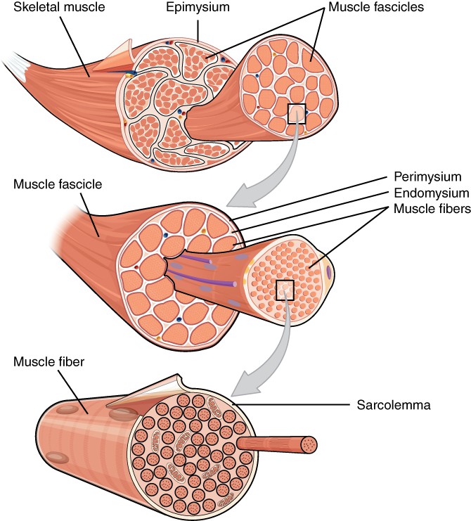
Figure 10.3 The Three Connective Tissue Layers Bundles of muscle fibres, called fascicles, are covered by the perimysium. Muscle fibres are covered by the endomysium.
Key Terms
actin protein that makes up most of the thin myofilaments in a sarcomere muscle fiber
action potential change in voltage of a cell membrane in response to a stimulus that results in transmission of an electrical signal; unique to neurons and muscle fibers
aponeurosis broad, tendon-like sheet of connective tissue that attaches a skeletal muscle to another skeletal muscle or to a bone
autorhythmicity heart’s ability to control its own contractions
cardiac muscle striated muscle found in the heart; joined to one another at intercalated discs and under the regulation of pacemaker cells, which contract as one unit to pump blood through the circulatory system. Cardiac muscle is under involuntary control.
contractility ability to shorten (contract) forcibly
desmosome cell structure that anchors the ends of cardiac muscle fibers to allow contraction to occur
eccentric contraction muscle contraction that lengthens the muscle as the tension is diminished
elasticity ability to stretch and rebound
endomysium loose, and well-hydrated connective tissue covering each muscle fiber in a skeletal muscle
epimysium outer layer of connective tissue around a skeletal muscle
excitability ability to undergo neural stimulation
extensibility ability to lengthen (extend)
fascicle bundle of muscle fibers within a skeletal muscle
intercalated disc part of the sarcolemma that connects cardiac tissue, and contains gap junctions and desmosomes
myoblast muscle-forming stem cell
myofibril long, cylindrical organelle that runs parallel within the muscle fiber and contains the sarcomeres
myosin protein that makes up most of the thick cylindrical myofilament within a sarcomere muscle fiber
perimysium connective tissue that bundles skeletal muscle fibers into fascicles within a skeletal muscle
skeletal muscle striated, multinucleated muscle that requires signaling from the nervous system to trigger contraction; most skeletal muscles are referred to as voluntary muscles that move bones and produce movement
smooth muscle nonstriated, mononucleated muscle in the skin that is associated with hair follicles; assists in moving materials in the walls of internal organs, blood vessels, and internal passageways
