
Chapter 13: THE SENSES
Introduction
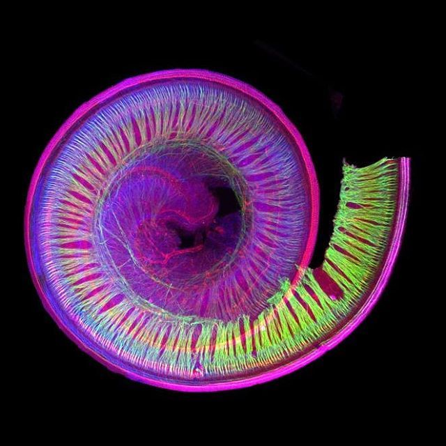
Figure 14.1 This microscopic view of the human inner ear explains why this sensory organ is called the cochlea (Gk. snail). Note the spiral shape of this organ, similar to the shell of a snail. [https://www.genengnews.com/insights/targeting-the-inner-ear/]
Chapter Objectives
After studying this chapter, you will be able to:
- Distinguish between general and special senses
- Describe different types of sensory receptors
- Describe and identify the major structures responsible for the special senses of taste, smell, hearing, balance, and vision
- Describe the different types of sensory receptors associated with somatosensation
- Describe the course of sensory information between the peripheral and central nervous system for reflex movement and cortical processing
A major role of sensory receptors is to help us learn about the environment around us, or about the state of our internal environment. Stimuli from varying sources, and of different types, are received and changed into the electrochemical signals of the nervous system. The sensory cell therefore relays the stimulus information to the central nervous system (CNS), where it is integrated with other sensory information—or sometimes higher cognitive functions—to become a conscious perception of that stimulus. The central integration may then lead to a motor response.
Describing sensory function with the term sensation or perception is a deliberate distinction. Sensation is the activation of sensory receptor cells at the level of the stimulus. Perception is the central processing of sensory stimuli into a meaningful pattern. Perception is dependent on sensation, but not all sensations are perceived. Receptors are the cells or structures that detect sensations.
Sensory Receptors
Stimuli in the environment activate specialised receptor cells in the peripheral nervous system. Different types of stimuli are sensed by different types of receptor cells. Receptor cells can be classified into types on the basis of three different criteria: cell type, position, and function.
Structural Receptor Types
The cells that interpret information about the environment can be either (1) a neuron that has a free nerve ending, with dendrites embedded in tissue that would receive a sensation; (2) a neuron that has an encapsulated ending in which the sensory nerve endings are encapsulated in connective tissue that enhances their sensitivity; or (3) a specialised receptor cell, which has distinct structural components that interpret a specific type of stimulus (Figure 14.2). The pain and temperature receptors in the dermis of the skin are examples of neurons that have free nerve endings. Also located in the dermis of the skin are lamellated corpuscles, neurons with encapsulated nerve endings that respond to pressure and touch. The cells in the retina that respond to light stimuli are an example of a specialised receptor, a photoreceptor.
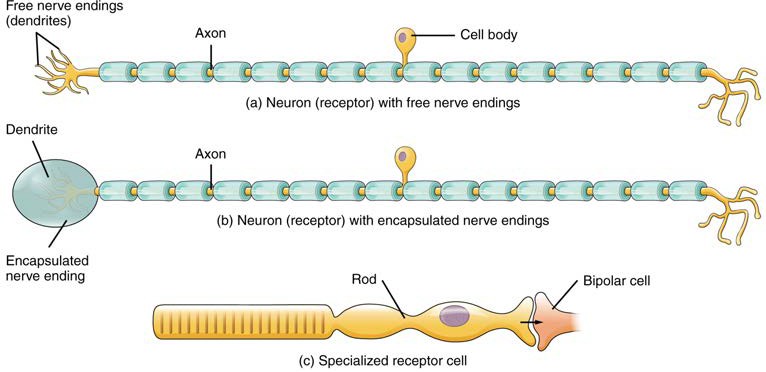
Figure 14.2 Receptor Classification by Cell Type Receptor cell types can be classified on the basis of their structure. Sensory neurons can have either (a) free nerve endings or (b) encapsulated endings. Photoreceptors in the eyes, such as rod cells, are examples of (c) specialised receptor cells.
Another way that receptors can be classified is based on their location relative to the stimuli. An exteroceptor is a receptor that is located near a stimulus in the external environment, such as the somatosensory receptors that are located in the skin. An interoceptor is one that interprets stimuli from internal organs and tissues, such as the receptors that sense the increase in blood pressure in the aorta or carotid sinus. Finally, a proprioceptor is a receptor located near a moving part of the body, such as a muscle, that interprets the positions of the tissues as they move.
Functional Receptor Types
Receptor cells can be further categorised on the basis of the type of stimuli they transduce. Chemical stimuli can be interpreted by a chemoreceptor that interprets chemical stimuli, such as an object’s taste or smell. Osmoreceptors respond to solute concentrations of body fluids. Additionally, pain is primarily a chemical sense that interprets the presence of chemicals from tissue damage, or similar intense stimuli, through a nociceptor. Physical stimuli, such as pressure and vibration, as well as the sensation of sound and body position (balance), are interpreted through a mechanoreceptor. Another physical stimulus that has its own type of receptor is temperature, which is sensed through a thermoreceptor that is either sensitive to temperatures above (heat) or below (cold) normal body temperature.
Special Senses
Senses can be classified as either general or specific. A general sense is one that is distributed throughout the body and has receptor cells within the structures of other organs. Mechanoreceptors in the skin, muscles, or the walls of blood vessels are examples of this type. General senses often contribute to the sense of touch, or to proprioception (touch) and kinesthesia (body movement), or to a visceral sense (organ position/movement), which is most important to autonomic functions. A special sense is one that has a specific organ devoted to it, namely the eye, inner ear, tongue, or nose.
Gustation (Taste)
Gustation is the special sense associated with the tongue. The surface of the tongue, along with the rest of the oral cavity, is lined by a stratified squamous epithelium. Raised bumps called papillae (singular = papilla) contain the structures for gustatory transduction. Within the structure of the papillae are taste buds that contain specialised gustatory receptor cells for the transduction of taste stimuli. These receptor cells are sensitive to the chemicals contained within foods that are ingested, and they release neurotransmitters based on the amount of the chemical in the food. Neurotransmitters from the gustatory cells can activate sensory neurons in the facial, glossopharyngeal, and vagus cranial nerves.
Olfaction (Smell)
Like taste, the sense of smell, or olfaction, is also responsive to chemical stimuli. The olfactory receptor neurons are located in a small region within the superior nasal cavity (Figure 14.4). This region is referred to as the olfactory epithelium and contains bipolar sensory neurons. Each olfactory sensory neuron has dendrites that extend from the apical surface of the epithelium into the mucous lining the cavity. As airborne molecules are inhaled through the nose, they pass over the olfactory epithelial region and dissolve into the mucous.
The axon of an olfactory neuron extends from the epithelium, through the floor of the cranial cavity to reach the brain. Olfactory neurons travel to multiple regions of the cerebrum, including the primary olfactory cortex that is located in the inferior and medial areas of the temporal lobe and the limbic system and hypothalamus, where smells become associated with long-term memory and emotional responses. This is how certain smells trigger emotional memories. Smell is the one sensory modality that does not synapse in the thalamus before connecting to the cerebral cortex. This intimate connection between the olfactory system and the cerebral cortex is one reason why smell can be a potent trigger of memories and emotion.
Audition (Hearing)
Hearing, or audition, is the transduction of sound waves into a neural signal that is made possible by the structures of the ear (Figure 14.5). The large, fleshy structure on the lateral aspect of the head is known as the auricle. The C-shaped curves of the auricle direct sound waves toward the external auditory canal (external acoustic meatus) of the temporal bone. At the end of the external auditory canal is the tympanic membrane, or eardrum, which vibrates after it is struck by sound waves. The auricle, external auditory canal, and tympanic membrane are often referred to as the external ear. The middle ear consists of a space spanned by three small bones called the middle ear ossicles. The three ossicles are the malleus, incus, and stapes, which are Latin names that roughly translate to hammer, anvil, and stirrup, respectively. The malleus is attached to the tympanic membrane and articulates with the incus. The incus, in turn, articulates with the stapes. The stapes is then attached to the inner ear, where the sound waves will be transduced into a neural signal. The middle ear is connected to the pharynx through the Eustachian tube (pharyngotympanic tube), which helps equilibrate air pressure across the tympanic membrane. The tube is normally closed but will pop open when the muscles of the pharynx contract during swallowing or yawning.
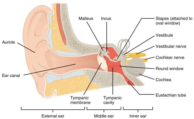
Figure 14.5 Structures of the Ear The external ear contains the auricle, external auditory canal and tympanic membrane. The middle ear contains the middle ear ossicles and is connected to the pharynx by the Eustachian tube. The inner ear contains the cochlea and vestibule, which are responsible for audition and equilibrium, respectively.
The inner ear is often described as a bony labyrinth, as it is composed of a series of canals embedded within the temporal bone. It has two separate regions, the cochlea and the vestibule, which are responsible for hearing and balance, respectively. The neural signals from these two regions are relayed to the brainstem through separate fibre bundles. However, these two distinct bundles travel together from the inner ear to the brainstem as the vestibulocochlear nerve. Sound is transduced into neural signals within the spiral-shaped cochlear region of the inner ear, which contains the sensory neurons in the Organ of Corti. The cochlea is connected to the stapes through a small ‘window’ such that sound transmitted through the auditory ossicles is transferred to the fluid filled chambers of the cochlea for sensory detection.
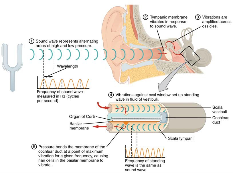
Figure 14.6 Transmission of Sound Waves to Cochlea A sound wave causes the tympanic membrane to vibrate. This vibration is amplified as it moves across the malleus, incus and stapes. The amplified vibration is picked up by a window at the boundary between the middle and inner ear causing pressure waves in the fluid of the inner ear. The complexity of the pressure waves is determined by the changes in amplitude and frequency of the sound waves entering the ear.
The organs of Corti contain hair cells, which are named for the hair-like stereocilia extending from the cell’s apical surfaces. The stereocilia are an array of microvilli-like structures arranged from tallest to shortest. The movement of the sterocilia results in signal transduction of the sensory receptors and activation of the cochlear branch of the vestibulocochlear nerve to initiate sound information to travel to the cerebrum.
INTERACTIVE ACTIVITY
Now review photos of the ear plastic model by clicking the arrows beside each image, or the buttons below the image panel. Each panel shows a different view after removal of further layers and parts of the model to identify the deeper structures. Test your knowledge on the unlabelled photos before revealing the names of the main features in the labelled photo. [Created in Anatomy.TV, Primal Pictures]
Questions:
(a) What is the importance of the Eustacian tube?
(b) What is the sensory function of the cochlea compared to the vestibule of the inner ear?
INTERACTIVE ACTIVITY
Equilibrium (Balance)
Along with audition, the inner ear is responsible for encoding information about equilibrium, the sense of balance. A similar mechanoreceptor—a hair cell with stereocilia—senses head position, head movement, and whether our bodies are in motion. These cells are located within the vestibule of the inner ear. Head position is sensed by the utricle and saccule, whereas head movement is sensed by the semicircular canals. The neural signals generated in the vestibule are transmitted through the vestibulocochlear nerve to the brainstem and cerebellum.
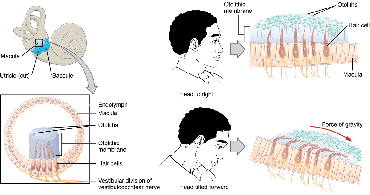
Figure 14.11 Linear Acceleration Coding in Utricle and Saccule of Vestibule Specialised receptors in the vestibule sense linear acceleration, such as when gravity acts on the tilting head, or if the head starts moving in a straight line.
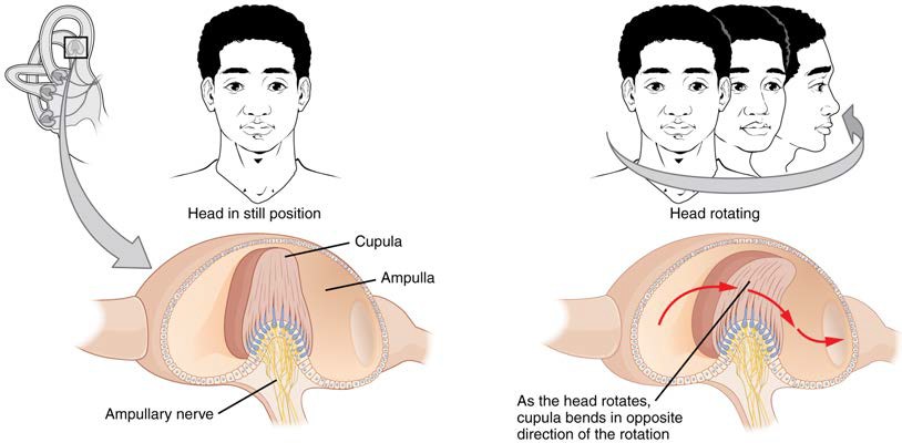
Figure 14.12 Rotational Coding by Semicircular Canals of Vestibule Rotational movement of the head is encoded by the hair cells in the base of the semicircular canals. As one of the canals moves in an arc with the head, the internal fluid moves in the opposite direction, causing the stereocilia to bend. The movement within the canals results in information about the direction in which the head is moving, and can give a very precise indication of head movement in three dimensions.
Vision
Vision is the special sense of sight that is based on the transduction of light stimuli received through the eyes. The eyes are located within either orbit in the skull. The bony orbits surround the eyeballs, protecting them and anchoring the soft tissues of the eye (Figure 14.13). The eyelids, with lashes at their leading edges, help to protect the eye from abrasions by blocking particles that may land on the surface of the eye. The inner surface of each lid is a thin membrane known as the palpebral conjunctiva. The conjunctiva extends over the white areas of the eye (the sclera), connecting the eyelids to the eyeball. Tears are produced by the lacrimal gland, located deep to the lateral edges of the nose. Tears produced by this gland flow through the lacrimal duct to the medial corner of the eye, where the tears flow over the eye, washing away foreign particles.
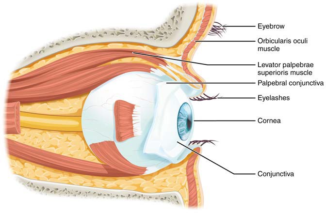
Figure 14.13 The Eye in the Orbit The eye is located within the orbit and surrounded by soft tissues that protect and support its function. The orbit is surrounded by cranial bones of the skull.
Movement of the eye within the orbit is accomplished by the contraction of six extraocular muscles that originate from the bones of the orbit and insert into the surface of the eyeball (Figure 14.14). Four of the muscles are arranged at the cardinal points around the eye and are named for those locations. They are the superior rectus, medial rectus, inferior rectus, and lateral rectus. When each of these muscles contract, the eye moves toward the contracting muscle. For example, when the superior rectus contracts, the eye rotates to look up. The superior oblique originates at the posterior orbit, near the origin of the four rectus muscles. However, the tendon of the oblique muscles threads through a pulley-like piece of cartilage known as the trochlea. The tendon inserts obliquely into the superior surface of the eye. The angle of the tendon through the trochlea means that contraction of the superior oblique rotates the eye medially. The inferior oblique muscle originates from the floor of the orbit and inserts into the inferolateral surface of the eye. When it contracts, it laterally rotates the eye, in opposition to the superior oblique (see Figure 14.13).
The extraocular muscles are innervated by three cranial nerves. The lateral rectus, which causes abduction of the eye, is innervated by the abducens nerve. The superior oblique is innervated by the trochlear nerve. All of the other muscles are innervated by the oculomotor nerve. The motor nuclei of these cranial nerves connect to the brainstem, which coordinates eye movements.
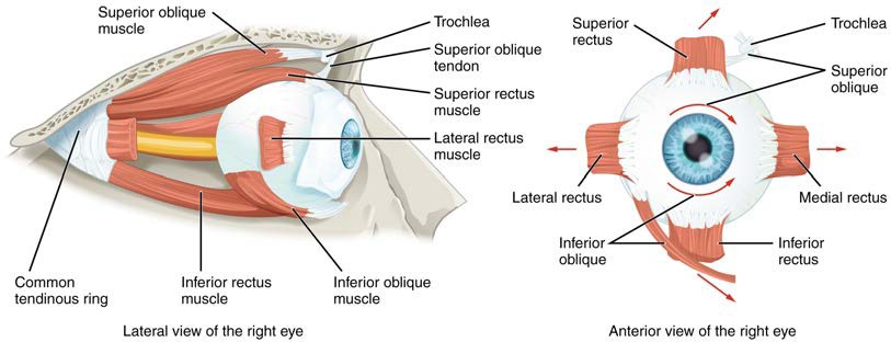
Figure 14.14 Extraocular Muscles The extraocular muscles move the eye within the orbit.
The eye itself is a hollow sphere composed of three layers of tissue: the fibrous tunic, the vascular tunic and the neural tunic (Figure 14.15).
The eye is also divided into two cavities: the anterior cavity (aqueous chamber) and the posterior cavity (vitreous chamber). The anterior cavity is the space between the cornea and lens, including the iris and ciliary body. It is filled with a watery fluid called the aqueous humor. The posterior cavity is the space posterior to the lens that extends to the posterior side of the interior eyeball, where the retina is located. The posterior cavity is filled with a more viscous fluid called the vitreous humor.
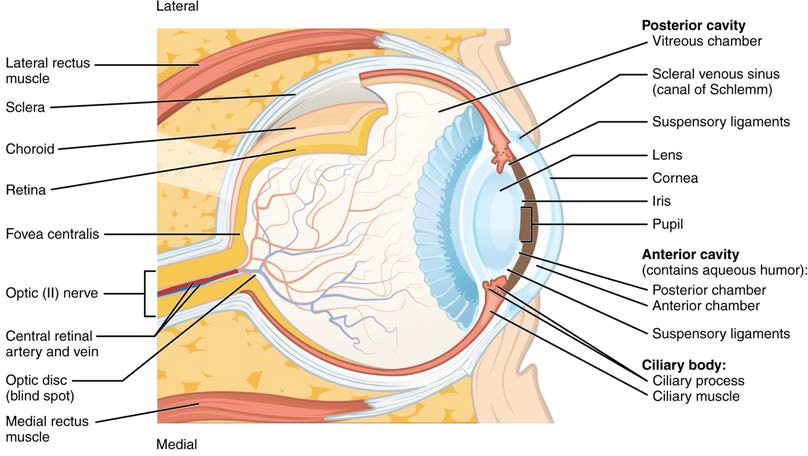
Figure 14.15 Structure of the Eye The sphere of the eye can be divided into anterior and posterior chambers. The wall of the eye is composed of three layers: the fibrous tunic, vascular tunic, and neural tunic.
INTERACTIVE ACTIVITY
Now review photos of the eye plastic model by clicking the arrows beside each image, or the buttons below the image panel. Each panel shows a different view after removal of further layers and parts of the model to identify the deeper structures. Test your knowledge on the unlabelled photos before revealing the names of the main features in the labelled photo. [Created in Anatomy.TV, Primal Pictures]
Question:
(a) Which structures are part of the fibrous, vascular and neural tunics?
INTERACTIVE ACTIVITY
This video from the QUT anatomy laboratory (2:35 minutes) demonstrates the anatomy of the fibrous, vascular and sensory tunics of the eye. [Holly Peters, Queensland University of Technology]
General Senses or Somatosensation
Somatosensation is considered a general sense, as opposed to the special senses discussed earlier. Somatosensation is the group of sensory modalities that are associated with touch, proprioception and interoception. These modalities include pressure, vibration, light touch, tickle, itch, temperature, pain, proprioception, and kinesthesia. This means that its receptors are not associated with a specialised organ, but are instead spread throughout the body in a variety of organs. Many of the somatosensory receptors are located in the skin, but receptors are also found in muscles, tendons, joint capsules, ligaments, and in the walls of visceral organs.
Two types of somatosensory signals that are transduced by free nerve endings are pain and temperature. These two modalities use thermoreceptors and nociceptors to transduce temperature and pain stimuli, respectively. Temperature receptors are stimulated when local temperatures differ from body temperature. Some thermoreceptors are sensitive to just cold and others to just heat. Nociception is the sensation of potentially damaging stimuli. Mechanical, chemical, or thermal stimuli beyond a set threshold will elicit painful sensations. Stressed or damaged tissues release chemicals that activate receptor proteins in the nociceptors.
If you drag your finger across a textured surface, the skin of your finger will vibrate. Such low frequency vibrations are sensed by mechanoreceptors called Merkel cells, also known as type I cutaneous mechanoreceptors. Merkel cells are located in the stratum basale of the epidermis. Deep pressure and vibration is transduced by lamellated (Pacinian) corpuscles, which are receptors with encapsulated endings found deep in the dermis, or subcutaneous tissue. Light touch is transduced by the encapsulated endings known as tactile (Meissner) corpuscles. Follicles are also wrapped in a plexus of nerve endings known as the hair follicle plexus. These nerve endings detect the movement of hair at the surface of the skin, such as when an insect may be walking along the skin. Stretching of the skin is transduced by stretch receptors known as bulbous corpuscles. Bulbous corpuscles are also known as Ruffini corpuscles, or type II cutaneous mechanoreceptors.
Other somatosensory receptors are found in the joints and muscles. Stretch receptors monitor the stretching of tendons, muscles and the components of joints. For example, have you ever stretched your muscles before or after exercise and noticed that you can only stretch so far before your muscles spasm back to a less stretched state? This spasm is a reflex that is initiated by stretch receptors to avoid muscle tearing. Such stretch receptors can also prevent over-contraction of a muscle. In skeletal muscle tissue, these stretch receptors are called muscle spindles. Golgi tendon organs similarly transduce the stretch levels of tendons. Bulbous corpuscles are also present in joint capsules, where they measure stretch in the components of the skeletal system within the joint. The types of nerve endings, their locations and the stimuli they transduce are presented in Table 14.1.
Table 14.1 Mechanoreceptors of Somatosensation
| Name | Historical (eponymous) | Location(s) | Stimuli |
| Free nerve endings | * | Dermis, cornea, tongue, joint capsules, visceral organs | Pain, temperature, mechanical deformation |
| Mechanoreceptors | Merkel’s discs | Epidermal-dermal junction, mucosal membranes | Low frequency vibrations (5-15 Hz) |
| Bulbous corpuscle | Ruffini’s corpuscle | Dermis, joint capsules | Stretch |
| Tactile corpuscle | Meissner’s corpuscle | Papillary dermis, especially in the fingertips and lips | Light touch, vibrations below 50 Hz |
| Lamellated corpuscle | Pacinian corpuscle | Deep dermis, subcutaneous tissue | Deep pressure, high-frequency vibration (around 250 Hz) |
| Hair follicle plexus | * | Wrapped around hair follicles in the dermis | Movement of hair |
| Muscle spindle | * | In line with skeletal muscle fibers | Muscle contraction and stretch |
| Tendon stretch organ | Golgi tendon organ | In line with tendons | Stretch of tendons |
Sensory Pathways
Specific regions of the CNS coordinate different movements of the body using sensory inputs and motor outputs of peripheral nerves. A simple case is a reflex caused by a synapse between a dorsal horn sensory neuron axon and a motor neuron in the ventral horn of the spinal cord. More complex arrangements are possible to integrate peripheral sensory information with higher processes. The important regions of the CNS that play a role in somatic processes can be separated into the spinal cord, brainstem, diencephalon, cerebral cortex and subcortical structures.
Spinal Nerves
Generally, spinal nerves contain afferent axons from sensory receptors in the periphery, such as from the skin, mixed with efferent axons travelling to the muscles or other effector organs. As the spinal nerve nears the spinal cord, it splits into dorsal and ventral roots. The dorsal root contains only the axons of sensory neurons, whereas the ventral roots contain only the axons of the motor neurons. Typically, spinal nerve systems that connect to the brain are contralateral, in that the right side of the body is connected to the left side of the brain and the left side of the body to the right side of the brain.
Cranial Nerves
Cranial nerves convey specific sensory information from the head and neck directly to the brain. For sensations inferior to the neck, the right side of the body is connected to the left side of the brain and the left side of the body to the right side of the brain. Whereas spinal information is contralateral, cranial nerve systems are mostly ipsilateral, meaning that a cranial nerve on the right side of the head is connected to the right side of the brain. Some cranial nerves contain only sensory axons, such as the olfactory, optic, and vestibulocochlear nerves. Other cranial nerves contain both sensory and motor axons, including the trigeminal, facial and vagus nerves. The general senses of somatosensation for the face travel through the trigeminal system.
Spinal cord pathways and cerebral integration of sensory information
A sensory pathway that carries peripheral sensations to the brain is referred to as an ascending tract. The various sensory modalities each follow specific pathways through the CNS. An example of an ascending tract is the spinothalamic tract that is primarily responsible for pain and temperature sensations. The name “spinothalamic” comes from the pathway of neurons between the spinal cord and contralateral (opposite side) thalamus to then travel to the postcentral gyrus of the parietal lobe of the cerebral cortex (primary somatosensory cortex).
In the cerebral cortex, the initial processing of sensory perception progresses to associative processing and then integration in multimodal areas of the cortex. These levels of processing can lead to the incorporation of sensory perceptions into memory, but more importantly, they lead to a response. In some cases this may elicit a motor response via neurons travelling through descending tracts through the CNS such as the corticospinal tract. The corticospinal tract contains axons of motor neurons that have their cell bodies housed in the precentral gyrus of the frontal lobe of the cerebral cortex. Their axons then pass to the contralateral side of the spinal cord where it synapses with a second neuron that travels to the skeletal muscle to initiate muscle contraction.
Reflexes
Simple somatic reflexes do not include the higher centres discussed for conscious or voluntary aspects of movement. Reflexes can be spinal or cranial, depending on the nerves and central components that are involved. A example of a somatic reflex is the withdrawal reflex to painful stimuli such as your hand touching a hot stove. This automatic and rapid withdrawal of your hand occurs through a series of three neurons that form a loop through the spinal cord. Nociceptors in the skin of the hand send sensory information via a sensory neuron to the dorsal horn of the spinal cord where it connects with multiple interneurons that synapse with motor neurons housed in the ventral horn of the spinal cord. These motor neuron then activate the biceps brachii muscle to flex the elbow and inhibit the antagonist triceps brachii muscle to remove the hand safely from the hot stove.
INTERACTIVE LINK
Another type of reflex is a stretch reflex. In this reflex, when a skeletal muscle is stretched, a muscle spindle receptor is activated. The axon from this receptor structure will cause direct contraction of the muscle. A collateral of the muscle spindle fibre will also inhibit the motor neuron of the antagonist muscles. The reflex helps to maintain muscles at a constant length. A common example of this reflex is the knee jerk that is elicited by a rubber hammer struck against the patellar ligament in a clinical exam.
Key Terms
aqueous humor watery fluid that fills the anterior chamber containing the cornea, iris, ciliary body, and lens of the eye
ascending tract fibre structure that relays sensory information from the periphery through the spinal cord and brain stem to other structures of the brain
association area region of cortex connected to a primary sensory cortical area that further processes the information to generate more complex sensory perceptions
audition sense of hearing
auditory ossicles three small bones in the middle ear
auricle fleshy external structure of the ear
chemoreceptor sensory receptor cell that is sensitive to chemical stimuli, such as in taste, smell, or pain
choroid highly vascular tissue in the wall of the eye that supplies the outer retina with blood
ciliary body smooth muscle structure on the interior surface of the iris that controls the shape of the lens through the zonule fibers
cochlea auditory portion of the inner ear containing structures to transduce sound stimuli
contralateral word meaning “on the opposite side,” as in axons that cross the midline in a fiber tract
cornea fibrous covering of the anterior region of the eye that is transparent so that light can pass through it
corticospinal tract connection between the cortex and the spinal cord responsible for generating movement
encapsulated ending configuration of a sensory receptor neuron with dendrites surrounded by specialized structures to aid in transduction of a particular type of sensation, such as the lamellated corpuscles in the deep dermis and subcutaneous tissue
equilibrium sense of balance that includes sensations of position and movement of the head
external ear structures on the lateral surface of the head, including the auricle and the ear canal back to the tympanic membrane
extraocular muscle one of six muscles originating out of the bones of the orbit and inserting into the surface of the eye which are responsible for moving the eye
fibrous tunic outer layer of the eye primarily composed of connective tissue known as the sclera and cornea
free nerve ending configuration of a sensory receptor neuron with dendrites in the connective tissue of the organ, such as in the dermis of the skin, that are most often sensitive to chemical, thermal, and mechanical stimuli
general sense any sensory system that is distributed throughout the body and incorporated into organs of multiple other systems, such as the walls of the digestive organs or the skin
gustation sense of taste
gustatory receptor cells sensory cells in the taste bud that transduce the chemical stimuli of gustation
hair cells mechanoreceptor cells found in the inner ear that transduce stimuli for the senses of hearing and balance
incus ossicle of the middle ear that connects the malleus to the stapes
inferior oblique extraocular muscle responsible for lateral rotation of the eye
inferior rectus extraocular muscle responsible for looking down
inner ear structure within the temporal bone that contains the sensory apparati of hearing and balance
interoceptor sensory receptor that is positioned to interpret stimuli from internal organs, such as stretch receptors in the wall of blood vessels
ipsilateral word meaning on the same side, as in axons that do not cross the midline in a fiber tract
iris colored portion of the anterior eye that surrounds the pupil
kinesthesia sense of body movement based on sensations in skeletal muscles, tendons, joints, and the skin
lacrimal duct duct in the medial corner of the orbit that drains tears into the nasal cavity
lacrimal gland gland lateral to the orbit that produces tears to wash across the surface of the eye
lateral rectus extraocular muscle responsible for abduction of the eye
lens component of the eye that focuses light on the retina
malleus ossicle that is directly attached to the tympanic membrane
mechanoreceptor receptor cell that transduces mechanical stimuli into an electrochemical signal
medial rectus extraocular muscle responsible for adduction of the eye
middle ear space within the temporal bone between the ear canal and bony labyrinth where the ossicles amplify sound waves from the tympanic membrane to the oval window
neural tunic layer of the eye that contains nervous tissue, namely the retina
nociceptor receptor cell that senses pain stimuli
olfaction sense of smell
olfactory epithelium region of the nasal epithelium where olfactory neurons are located
olfactory sensory neuron receptor cell of the olfactory system, sensitive to the chemical stimuli of smell, the axons of which compose the first cranial nerve
optic nerve second cranial nerve, which is responsible visual sensation
organ of Corti structure in the cochlea in which hair cells transduce movements from sound waves into electrochemical signals
papilla for gustation, a bump-like projection on the surface of the tongue that contains taste buds
photoreceptor receptor cell specialized to respond to light stimuli
primary somatosensory cortex region of the cerebral cortex that initially receives sensory input from an ascending pathway from the thalamus and begins the processing that will result in conscious perception of that modality
proprioception sense of position and movement of the body
proprioceptor receptor cell that senses changes in the position and kinesthetic aspects of the body
pupil open hole at the center of the iris that light passes through into the eye
receptor cell cell that transduces environmental stimuli into neural signals
retina nervous tissue of the eye at which phototransduction takes place
sclera white of the eye
sensory modality a particular system for interpreting and perceiving environmental stimuli by the nervous system
somatosensation general sense associated with modalities lumped together as touch
special sense any sensory system associated with a specific organ structure, namely smell, taste, sight, hearing, and balance
spinothalamic tract ascending tract of the spinal cord associated with pain and temperature sensations
stapes ossicle of the middle ear that is attached to the inner ear
stereocilia array of apical membrane extensions in a hair cell that transduce movements when they are bent
stretch reflex response to activation of the muscle spindle stretch receptor that causes contraction of the muscle to maintain a constant length
superior oblique extraocular muscle responsible for medial rotation of the eye
superior rectus extraocular muscle responsible for looking up
taste buds structures within a papilla on the tongue that contain gustatory receptor cells
thermoreceptor sensory receptor specialized for temperature stimuli
trochlea cartilaginous structure that acts like a pulley for the superior oblique muscle
tympanic membrane ear drum
vascular tunic middle layer of the eye primarily composed of connective tissue with a rich blood supply
visceral sense sense associated with the internal organs
vision special sense of sight based on transduction of light stimuli
vitreous humor viscous fluid that fills the posterior chamber of the eye
zonule fibers fibrous connections between the ciliary body and the lens