
Digital Capabilities Case Study 1
Digital Capabilities Case Study: LQB670 Anatomical Dissection
Organisational area: School of Biomedical Sciences, Faculty of Health, Queensland University of Technology, Australia
Course: Bachelor of Biomedical Science
Student cohort: 3rd year Biomedical Science students; capstone unit
QUT Assessment type: Case Study
Course Learning Outcomes mapped to case study assessment:
-
Critically review, analyse and synthesise foundational knowledge in a broad range of biomedical discipline areas and in depth theoretical, technical and practical knowledge in specialised discipline areas.
-
Demonstrate the cognitive skills required to find solutions to scientific problems.
-
Apply knowledge and skills to rapidly source, critically analyse and communicate biomedical science information using appropriate technologies.
Step 1: Identify relevant Digital Capabilities to embed
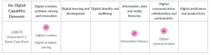
Having prioritised digital capability graduate needs of your student cohort, digital capability development has been scaffolded across the course and mapped to specific core units of study. As a capstone unit, a number of digital capabilities were mapped to the assessment in LQB670 across three Jisc elements. Digital capability subelements that requiring embedding in LQB670 are illustrated in the below table.
Step 2: Refine your Unit Learning Outcomes (ULO)
Having identified the digital capabilities that need to be developed and assessed in the unit, the unit learning outcomes should be reviewed to ensure the relevant digital capabilities are explicitly expressed in these outcomes. Review the refinements made to the ULOs in the example below for the Case Study assessment in LQB670: Anatomical Dissection.
LQB670 Anatomical Dissection Unit Learning Outcomes for Case Study Assessment
This assessment is mapped to Unit Learning Outcomes (ULO) 1 and ULO4.
ULO1
Use the process of dissection and computed tomography image interpretation to investigate and critically analyse clinical relationships between anatomical structures and pathology.
Revised: Use the process of dissection and digital problem-solving to investigate and critically analyse clinical relationships between anatomical structures and pathology to create scientific figures.
ULO4
Interrogate the literature to form an evidence-based opinion on the phenotypic range of anatomical variation within a population and hypothesise probable causes.
Revised: Interrogate the literature to form and digitally communicate an evidence-based opinion on the phenotypic range of anatomical variation within a population and hypothesise probable causes.
Step 3: Design your Assessment
The case study assessment type provides an authentic project-based setting to develop all four of the identified digital capabilities.
Mapping the Digital Capability elements in the assessment
Refer to the unit outline description of the assessment below and note how the digital capability subelements are embedded in the assessment design by referring to each footnote.
You will investigate and document the phenotypic expression of a range of anatomical variation throughout your donor body1 and research the population frequency of each phenotype2. From this list you will then select one organ to write an anatomical variation case report suitable for publication in the Open Educational Resource (OER)3 titled Anatomical Variation: An Australian and New Zealand Context. This report will describe the anatomical appearance of the organ in the donor and compare to other phenotypes documented in primary literature. The clinical impact and possible causes of anatomical variation in this organ structure will also be explored2. This OER will be made available to anatomy students across Australia and New Zealand to improve their understanding of anatomical variation.
Your case study will also include a detailed investigation of the major pathology visible in your donor body through macroscopic and computed tomography interpretation, to document the anatomical appearance of this pathology and estimate the cause of death of the donor4.
Incorporating the JISC Building Capabilities Framework into the assessment
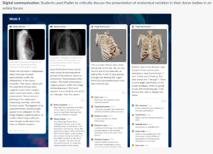
1Digital communication. Students use a Padlet to digitally communicate with their peers on their preliminary research findings on the anatomical variation they have identified. Through meaningful conversations students develop a deeper understanding of the breadth of anatomical variation in the human body. Students demonstrate respectful and professional communication approaches to support peer collaboration and the secure use of sensitive digital content (donor photos).
2Information literacy. As students are researching anatomical variation and pathological presentation, they are demonstrating their ability to find, evaluate, organise and share information for learning and research purposes using primary and secondary literature.
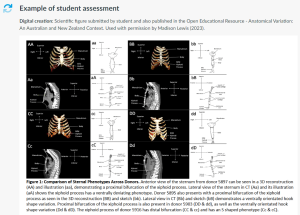
3Digital creation: Students create a scientific figure suitable for journal publication and the Open Educational Resource (OER) Pressbook, Anatomical Variation: An Australian and New Zealand Context.
4Digital problem-solving: As students need to investigate and determine the cause of death in the donor they will be demonstrating the ability to solve problems and make decisions on the likely pathologies present in their donor. In particular students are required to build specialist technical knowledge in the use of DICOM viewers to analyse and manipulate computed tomography scans in their evaluation of pathology in the donor body. You will also be encouraged to use Generative Artificial Intelligence to problem solve possible pathologies based on their anatomical observations.
Rubric to assess digital capability elements
Standards have been written to assess digital creation, digital problem-solving, digital communication and information literacy.
DIGITAL COMMUNICATION ON ANATOMICAL VARIATION (PADLET) (5%)
| 7 | · Digital posts utilise respectful and professional language and treatment of donor material.
· Digital communication with peers utilises respectful and constructive language, which encourages participation and creates a safe and inclusive digital learning environment. · Relevant, analytically complex and regular contributions to Padlet, which always promote critical thinking and deepen understanding of anatomical variation through extended discussion via prompting. |
| 6 | · Digital posts utilise respectful and professional language and treatment of donor material.
· Digital communication with peers utilises respectful and constructive language, which encourages participation and creates a safe and inclusive digital learning environment. · Relevant, analytical and regular contributions to Padlet, which always promote critical thinking and deepen understanding of anatomical variation through extended discussion. |
| 5 | · Digital posts utilise respectful and professional language and treatment of donor material.
· Digital communication with peers utilises respectful and constructive language, which creates a safe and inclusive digital learning environment. · Relevant, analytical and regular contributions to Padlet, which often promote critical thinking and deepen understanding of anatomical variation. |
| 4 | · Digital posts utilise respectful and professional language and treatment of donor material.
· Digital communication with peers utilises respectful language, which creates a safe and inclusive digital learning environment. · Relevant, analytical contributions to Padlet, which occasionally promote critical thinking and deepen understanding of anatomical variation. |
| 3 | · Digital posts mostly utilise respectful and professional language and treatment of donor material.
· Digital communication with peers mostly utilises respectful language, which creates a safe and inclusive digital learning environment. · Mostly relevant contributions to Padlet, which add superficial value to the discussion. |
| 2 | · Digital posts occasionally utilise respectful and professional language and treatment of donor material.
· Digital communication with peers occasionally utilises respectful language, which creates a safe and inclusive digital learning environment. · Frequently irrelevant or underdeveloped contributions to Padlet, which add little value to the discussion. |
| 1 | · Digital posts are disrespectful in language and treatment of donor material.
· Irrelevant or absent contributions to Padlet, which add no value to the discussion. |
DIGITAL CREATION OF SCIENTIFIC FIGURES (7.5%)
| 7 | · Uses secure processes to work with sensitive content in a digital environment.
· Careful selection and purposeful digital manipulation of images using a range of digital technologies enhances the visualisation of relevant anatomy to create figures suitable for scientific publication. · The anatomy in all figures is expertly communicated through accurate, sufficient and specific labels, headings and annotations using outstanding graphical design skills. |
| 6 | · Uses secure processes to work with sensitive content in a digital environment.
· Careful selection and digital manipulation of images enhances the visualisation of relevant anatomy to create figures suitable for scientific publication in most cases. · The anatomy in all figures is expertly communicated through accurate, sufficient and specific labels, headings and annotations using excellent graphical design skills. |
| 5 | · Uses secure processes to work with sensitive content in a digital environment.
· Careful selection and digital manipulation of images enhances the visualisation of relevant anatomy to create figures suitable for scientific publication in most cases. · The anatomy in figures is clearly communicated through accurate labels, headings and annotations using excellent graphical design skills. |
| 4 | · Uses secure processes to work with sensitive content in a digital environment.
· Careful selection and digital manipulation of images enhances the visualisation of anatomy to create figures suitable for scientific publication in most cases. · Essential information in figures is clearly communicated using basic graphical design skills. Anatomical structures have been accurately identified in most cases. |
| 3 | · Uses secure processes to work with sensitive content in a digital environment.
· Selection and digital manipulation of images is not the most appropriate or sufficient to clearly visualise the required anatomy. · Errors and insufficiency in anatomical identification results in superficial information on digital images which is difficult to interpret. |
| 2 | · Uses secure processes to work with sensitive content in a digital environment.
· Frequent errors in labelling and anatomical interpretation results in digital images that are not appropriate for publication. |
| 1 | · Does not use secure processes to work with sensitive content in a digital environment.
· Insufficient digital images to be able to visualise anatomical variation or pathology in the donor. |
CRITICAL ANALYSIS (10%)
| 7 | · Critical research skills are extensively used to investigate possible causes of phenotypic anatomical variation through a deep analysis of the effect of sex and population on phenotype incidence.
· Critical analysis of presentation, impact and relationships between the major pathologies identified in the donor is evidence-based to form a logical cause of death conclusion. |
| 6 | · Critical research skills are used to investigate possible causes of phenotypic anatomical variation through a deep analysis of the effect of sex and population on phenotype incidence.
· Critical analysis of presentation and impact between the major pathologies identified in the donor is evidence-based to form a logical cause of death conclusion, however it is not clear how the three pathologies link together and/or the order of presentation. |
| 5 | · Research skills are used to investigate possible causes of phenotypic anatomical variation through a detailed analysis of the effect of sex and population on phenotype incidence in most cases.
· Critical analysis of presentation and impact between the major pathologies identified in the donor is evidence-based to form a logical cause of death conclusion, however it is not clear how the three pathologies link together and/or the order of presentation. |
| 4 | · Basic research skills are used to investigate possible causes of phenotypic anatomical variation through a superficial analysis of the effect of sex and population on phenotype incidence that may be incomplete in a small number of occasions.
· Analysis of presentation, impact and relationships between the major pathologies identified in the donor forms a logical cause of death conclusion, however there is limited justification supported by literature. |
| 3 | · Basic research skills are used to investigate possible causes of phenotypic anatomical variation through an analysis of the effect of sex and population on phenotype incidence that contains instances of inappropriate or inaccurate descriptions.
· Analysis of presentation, impact and relationships between the major pathologies identified in the donor forms a logical cause of death conclusion, with limited justification supported by literature. |
| 2 | · Ineffective research skills are used to investigate possible causes of phenotypic anatomical variation through an analysis of the effect of sex and population on phenotype incidence that contains multiple instances of inappropriate or inaccurate descriptions or large incomplete sections.
· Analysis of presentation, impact and relationships between the major pathologies presents errors in interpretation resulting in flawed estimation of cause of death. |
| 1 | · Limited research skills are used to investigate possible causes of phenotypic anatomical variation with significant errors and/or limited information provided.
· Estimated cause of death presents major errors. |
ACADEMIC WRITING AND INFORMATION LITERACY (5%)
| 7 | · Writing adheres to the appropriate conventions of Australian/British spelling and grammar at all times and is organised in clear paragraphs.
· Contributions to digital communication and critical examination of variation and pathologies extensively use relevant research literature and evidence to develop and support ideas using accurate APA conventions and referencing. |
| 6 | · Writing adheres to the appropriate conventions of Australian/British spelling and grammar at all times and is organised in clear paragraphs.
· Contributions to digital communication and critical examination of variation and pathologies use research literature and evidence to develop and support ideas using accurate APA conventions and referencing. |
| 5 | · Minor errors in spelling and/or grammar. |
| 4 | · Minor errors in the reference list or in-text citations according to QUT APA referencing conventions. |
| 3 | · Limited research literature used to evidence anatomical description and ideas using APA conventions and referencing. |
| 2 | · Frequent errors in spelling, grammar, and/or referencing. Use of GenAI has not been appropriately acknowledged. |
| 1 | · No attempt to adhere to communication and referencing conventions results in a confusing response that may breach academic integrity expectations.
· No research literature evidenced. |
Synchronous Activities
Digital creation: workshop activity
- Students are asked to critique a range of high- and poor-quality scientific figures on the accuracy, interpretation and presentation of data as a whole of class discussion. Figures are drawn from authentic examples in the literature and academic-generated examples that align to the assessment task figure requirements. Live polling technology is used to collate student perspectives on each figure followed by academic-led discussion on the positive qualities of the figure and how the figure quality could be improved.
- Students are asked to create a scientific figure in Microsoft PowerPoint and upload to a class Padlet site for peer feedback and discussion.
- Academic leads a discussion on the marking rubric on digital creation to communicate assessment expectations and standards of achievement.
Digital problem-solving: workshop activity
- Students are asked to download a DICOM viewer and the DICOM files from a past year donor CT case to their device before class.
- Academic demonstrates how to use a DICOM viewer to analyse human computed tomography (CT) image datasets using live screencasting, encouraging students to follow along using their preferred DICOM viewer on their own device. Using an authentic radiologic approach, the academic guides the students on how to identify and diagnose pathologies in each part of the body in the CT images using the DICOM viewer.
- Academic discusses how Generative Artificial Intelligence (AI) may be used effectively to identify and diagnose pathologies. Ethical considerations and limitations of Generative AI are discussed.
- Students are asked to prompt their preferred Generative AI (Microsoft Copilot or ChatGPT used in academic examples) to list possible pathologies from an anatomical description provided by student on the CT appearance of a suspected pathology observed in the DICOM viewer. Academic leads a discussion on how to critically analyse each possible pathology identified by Generative AI using peer-reviewed literature.
Asynchronous Activities
Digital communication: Canvas guidance and resources
- Students are ask
Information literacy: Canvas guidance and resources
- Link to QUT Study Smart information research and literacy skills resource (QUT | Library | Study Smart | Home) added to Canvas. Students are recommended to complete Module 2: Find to review effective search strategies and selecting most appropriate tools for finding information and Module 3: Evaluate to assess the suitability of their search results and revise their search strategy if necessary.
- Link to QUT Cite|Write (QUT cite|write – QUT cite|write) added to Canvas. Students are recommended to review this resource to appropriately acknowledge source material using QUT APA referencing style.
